. 2006 Jan 1;90(1):287-97.
doi: 10.1529/biophysj.105.071035. Epub 2005 Oct 7.

Mechanical resistance of proteins explained using simple molecular models

Affiliations

Mechanical resistance of proteins explained using simple molecular models

Daniel K West et al. Biophys J. .

Abstract

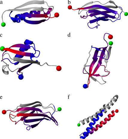
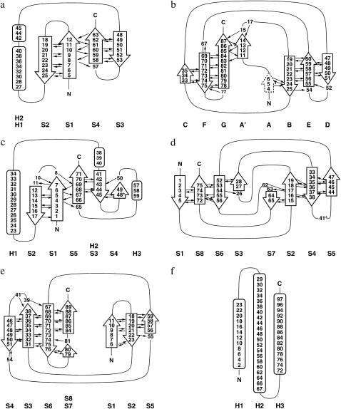
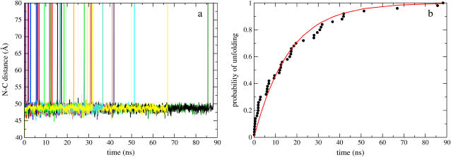
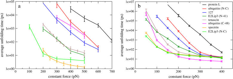
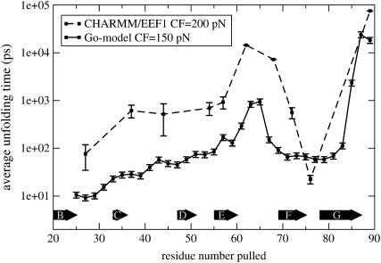
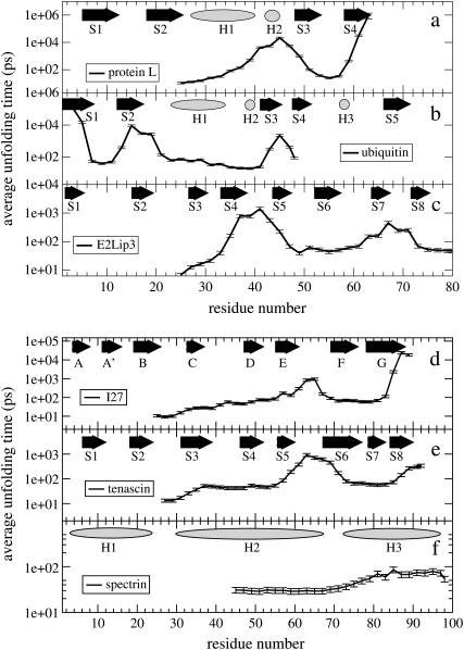
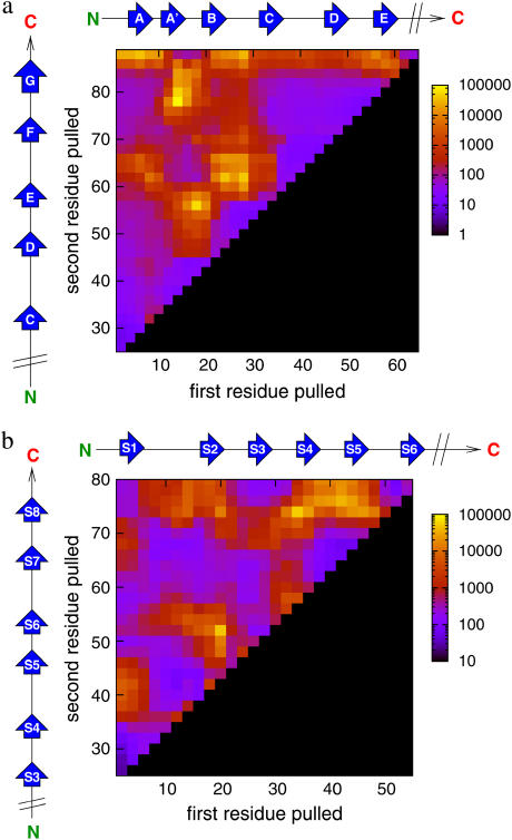
Recent experiments have demonstrated that proteins unfold when two atoms are mechanically pulled apart, and that this process is different to when heated or when a chemical denaturant is added to the solution. Experiments have also shown that the response of proteins to external forces is very diverse, some of them being "hard," and others "soft." Mechanical resistance originates from the presence of barriers on the energy landscape; together, experiment and simulation have demonstrated that unfolding occurs through alternative pathways when different pairs of atoms undergo mechanical extension. Here we use simulation to probe the mechanical resistance of six structurally diverse proteins when pulled in different directions. For this, we use two very different models: a detailed, transferable one, and a coarse-grained, structure-based one. The coarse-grained model gives results that are surprisingly similar to the detailed one and qualitatively agree with experiment; i.e., the mechanical resistance of different proteins or of a single protein pulled in different directions can be predicted by simulation. The results demonstrate the importance of pulling direction relative to the local topology in determining mechanical stability, and rationalize the effect of the location of importation/degradation tags on the rates of mitochondrial import or protein degradation in vivo.


Figures

FIGURE 1
FIGURE 1
Structure and mechanical strength of the six test proteins: (a) protein L, (b) I27, (c) ubiquitin, (d) E2lip3, (e) tenascin, and (f) spectrin. Blue to red represents soft to hard directions relative to that domain only when pulled from the N-terminus (green sphere), or C-terminus (red sphere) in the case of ubiquitin. Gray denotes regions not pulled. Experimentally E2lip3 and ubiquitin have been pulled from their N-/C-termini, and in a second direction, N-41 and C-48, respectively (denoted by the blue sphere). Figure generated using the program MOLMOL (54).
FIGURE 2
FIGURE 2
Topology diagrams of the six test proteins: (a) protein L, (b) I27, (c) ubiquitin, (d) E2lip3, (e) tenascin, and (f) spectrin. Black arrows indicate inter-β-strand hydrogen bonds.
FIGURE 3
FIGURE 3
(a) I27 N-C extension as a function of time when a constant 150 pN force is applied using the Gō model. A single unfolding barrier can clearly be seen at 49 ± 2 Å, from which unfolding occurs (N-C distance in the native state is 42 Å). (b) The probability of unfolding fits a single exponential formula image with τ0 = 16 ns.
FIGURE 4
FIGURE 4
The average unfolding time of six different protein domains under force pulled from their N-C termini using (a) a detailed, transferable EEF1 model and (b) a coarse-grained, structure-based Gō model. E2lip3 and ubiquitin are pulled in a second direction, revealing qualitative agreement with experiment. Note the broad range of forces over which the mechanical ranking of the proteins can be defined. The error bar for protein L at 150 pN in the Gō model has been removed for clarity.
FIGURE 5
FIGURE 5
The unfolding time profile of the detailed, transferable EEF1 model (CF = 200 pN) and coarse-grained, structure-based Gō model (CF = 150 pN), pulling I27 from the N-terminus and other Cα atoms within the domain.
FIGURE 6
FIGURE 6
Unfolding time as a function of the pair of Cα atoms to which the constant force (150 pN) is applied. In abscissa is the Cα atom to which force is applied, while the N-terminus is kept fixed (except for ubiquitin where the C-terminus is kept fixed). Along the top of each unfolding time profile is the native secondary structure; β-strands are shown as arrows, α-helices as ellipses.
FIGURE 7
FIGURE 7
Gō models reveal anisotropy in the mechanical unfolding landscape of (a) I27 (CF = 150 pN) and (b) E2lip3 (CF = 100 pN). Yellow to blue colors denote geometries of high to weak mechanical resistance, black denotes regions not pulled. Scale is in picoseconds.

References

    1. Rief, M., M. Gautel, F. Oesterhelt, J. M. Fernandez, and H. E. Gaub. 1997. Reversible unfolding of individual titin immunoglobulin domains by AFM. Science. 276:1109–1112. - PubMed
    1. Carrion-Vasquez, M., A. F. Oberhauser, S. B. Fowler, P. E. Marszalek, S. E. Broedel, J. Clarke, and J. M. Fernandez. 1999. Mechanical and chemical unfolding of a single protein: a comparison. Proc. Natl. Acad. Sci. USA. 96:3694–3699. - PMC - PubMed
    1. Fowler, S., R. B. Best, J. L. Toca-Herrera, T. Rutherford, A. Steward, E. Paci, M. Karplus, and J. Clarke. 2002. Mechanical unfolding of a titin Ig domain: structure of unfolding intermediate revealed by combining AFM, molecular dynamics simulations, NMR and protein engineering. J. Mol. Biol. 322:841–849. - PubMed
    1. Brockwell, D. J., E. Paci, R. C. Zinober, G. S. Beddard, P. D. Olmsted, D. A. Smith, R. N. Perham, and S. E. Radford. 2003. Pulling geometry defines the mechanical resistance of a β-sheet protein. Nat. Struct. Biol. 10:731–737. - PubMed
    1. Brockwell, D. J., G. S. Beddard, E. Paci, D. K. West, P. D. Olmsted, D. A. Smith, and S. E. Radford. 2005. Mechanically unfolding the small topologically simple protein L. Biophys. J. 89:506–519. - PMC - PubMed

Publication types

LinkOut - more resources