. 2005 Oct 18;102(42):15081-6.
doi: 10.1073/pnas.0502889102. Epub 2005 Oct 7.

Signal propagation from membrane messengers to nuclear effectors revealed by reporters of phosphoinositide dynamics and Akt activity

Affiliations

Signal propagation from membrane messengers to nuclear effectors revealed by reporters of phosphoinositide dynamics and Akt activity

Bharath Ananthanarayanan et al. Proc Natl Acad Sci U S A. .

Abstract

Among various second messengers, phosphatidylinositol 3,4,5-triphosphate (PIP3) and phosphatidylinositol 3,4-bisphosphate [PI(3,4)P2] regulate a variety of cellular processes, such as cell survival, polarization, and proliferation. Many of these functions are achieved via activation of serine/threonine kinase Akt. To investigate the spatiotemporal regulation of these lipids, we constructed a genetically targetable phosphoinositide (PI) indicator by sandwiching pleckstrin homology (PH) domain of Akt and a "pseudoligand" containing acidic amino acid residues, between cyan and yellow mutants of GFP. In living cells, elevations in PIP3 and PI(3,4)P2 by growth factor-induced activation of phosphatidylinositol 3-kinase (PI3K) resulted in a change in fluorescence resonance energy transfer (FRET) between the fluorescent proteins, increasing yellow to cyan emission ratios by 10-30%. This response can be reversed by inhibiting PI3K and abolished by mutating the critical residues responsible for PI binding. Differential dynamics of PIs were observed at plasma membrane of NIH 3T3 cells, stimulated by various growth factors. On the other hand, the nuclear targeted indicator showed no response within an hour after platelet-derived growth factor stimulation, suggesting that no appreciable amounts of accessible PIP3 and PI(3,4)P2 were produced in the nucleus. Furthermore, simultaneous imaging of a plasma membrane-targeted PI indicator and a nuclear-targeted Akt activity reporter revealed a gradual and sustained accumulation of Akt activity in the nucleus after rapid and transient production of PIP3 and PI(3,4)P2 at plasma membrane in the same cell. Thus, signal propagation from the lipid messengers at plasma membrane to the effectors in the nucleus is precisely controlled by kinases as well as lipid and protein phosphatases.


Figures

Fig. 1.
Fig. 1.
Development of InPAkt. (a) A cartoon depicting the conformational change upon PI binding, yielding a FRET response. (b) Domain structure of the construct showing the restriction sites linking individual components. (c) Anti-GFP Western blot of InPAkt expressed in HEK 293 cells indicating the right size of the reporter. (d) FRET response of InPAkt expressed in NIH 3T3 cells. Yellow fluorescence images show membrane translocation of InPAkt upon PDGF stimulation. Pseudocolor images indicate the emission ratio change at various time points after PDGF stimulation. (e) Representative emission ratio time courses of InPAkt (n = 9) and the PH domain mutant R23A/R25A (n = 2), both stimulated with 50 ng/ml PDGF. InPAkt showed a response of 25.4 ± 3.7% (average ± SD; n = 9). (f) A representative emission ratio time course from two independent experiments showing that the response of InPAkt is PI3K-specific. NIH 3T3 cells were pretreated with 20 μM PI3K inhibitor LY294002, followed by stimulation with 50 ng/ml PDGF, gentle washing, and treatment with PDGF again. (g) A representative emission ratio time course shows the reversibility of the reporter. NIH 3T3 cells expressing InPAkt were stimulated with PDGF, followed by treatment with 20 μM LY294002 (n = 5).
Fig. 2.
Fig. 2.
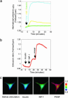
Comparison of the cellular responses stimulated by various growth factors. (a) Representative time courses of InPAkt responses in cells stimulated by 50 ng/ml PDGF (blue), 50 nM IGF-1 (yellow), and 100 ng/ml insulin (green) (n = 3). (b) Representative emission ratio time courses showing the FRET response of a NIH 3T3 cell expressing InPAkt sequentially stimulated by insulin, IGF-1, and PDGF (n = 3). (c) Pseudocolor images showing InPAkt responses with sequential stimulation by insulin, IGF-1, and PDGF.
Fig. 3.
Fig. 3.
Fusions of InPAkt targeted to various subcellular locations. (a) Domain structure of the fusion constructs. (b) Localization of plasma membrane targeted InPAkt (pm InPAkt) is shown in the fluorescence image (YFP). Pseudocolor images show colocalization of nuclear targeted reporter (NLS InPAkt) with a cell-permeable DNA dye, Hoechst 3342. (c) A representative emission ratio time course from four independent experiments showing the response of plasma membrane targeted InPAkt stimulated with 50 ng/ml PDGF (9.25 ± 0.4%), followed by addition of 20 μM LY294002. (d) A representative emission ratio time course from three different trials for NLS InPAkt in NIH 3T3 cells stimulated with PDGF.
Fig. 4.
Fig. 4.
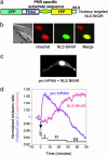
Simultaneous imaging of plasma membrane targeted InPAkt (pm InPAkt) and nuclear targeted BKAR (NLS BKAR). (a) Domain structure of NLS BKAR. (b) Pseudocolor images show colocalization of NLS BKAR with Hoechst 3342. (c) Cellular distribution of the two reporters in a HEK 293 cell. (d) Representative emission ratio time courses from four different experiments for pm InPAkt and NLS BKAR in the same cell stimulated with 50 nM IGF-1.

Similar articles

Cited by

References

    1. Martin, T. F. (1998) Annu. Rev. Cell Dev. Biol. 14, 231–264. - PubMed
    1. Toker, A. (2002) Cell Mol. Life Sci. 59, 761–779. - PMC - PubMed
    1. Fruman, D. A., Meyers, R. E. & Cantley, L. C. (1998) Annu. Rev. Biochem. 67, 481–507. - PubMed
    1. Maehama, T., Taylor, G. S. & Dixon, J. E. (2001) Annu. Rev. Biochem. 70, 247–279. - PubMed
    1. Krystal, G. (2000) Semin. Immunol. 12, 397–403. - PubMed

Publication types

MeSH terms

Substances