. 2005 Nov;17(11):2993-3006.
doi: 10.1105/tpc.105.036004. Epub 2005 Oct 7.

The NAC transcription factors NST1 and NST2 of Arabidopsis regulate secondary wall thickenings and are required for anther dehiscence

Affiliations

The NAC transcription factors NST1 and NST2 of Arabidopsis regulate secondary wall thickenings and are required for anther dehiscence

Nobutaka Mitsuda et al. Plant Cell. 2005 Nov.

Abstract

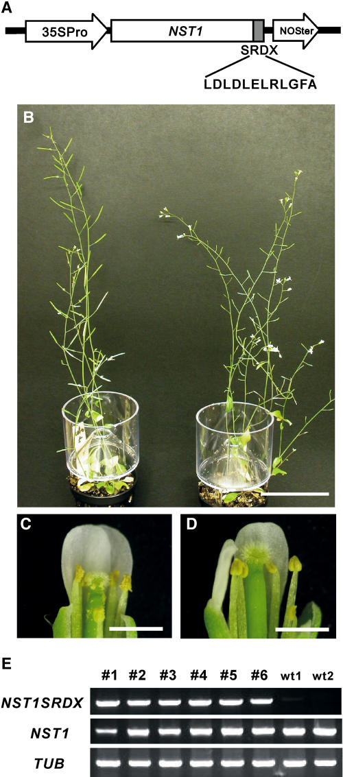
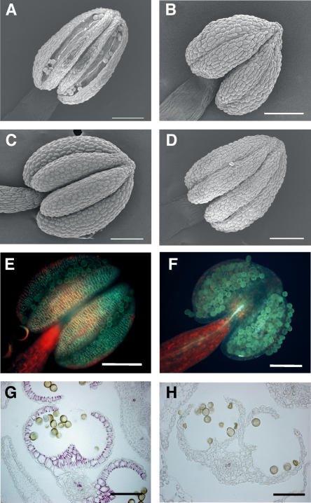
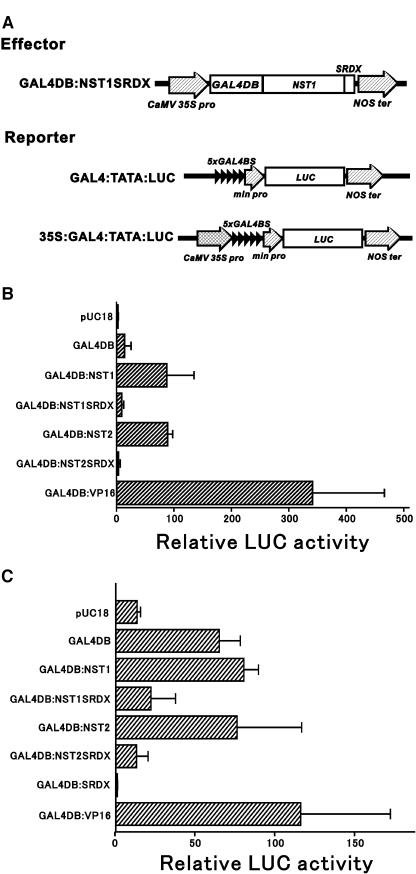
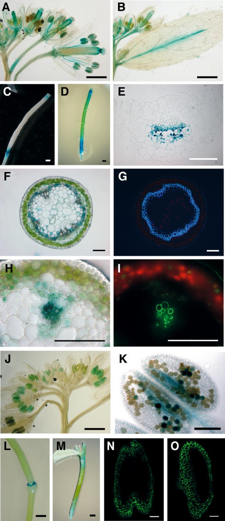
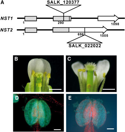
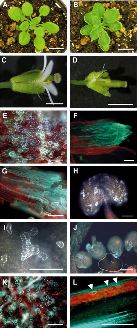
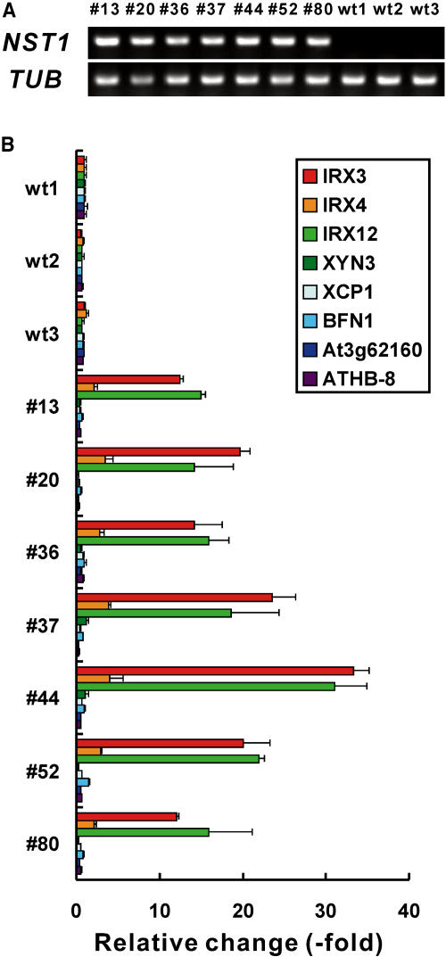
In plants, secondary wall thickenings play important roles in various biological processes, although the factors regulating these processes remain to be characterized. We show that expression of chimeric repressors derived from NAC SECONDARY WALL THICKENING PROMOTING FACTOR1 (NST1) and NST2 in Arabidopsis thaliana resulted in an anther dehiscence defect due to loss of secondary wall thickening in anther endothecium. Plants with double, but not single, T-DNA-tagged lines for NST1 and NST2 had the same anther-indehiscent phenotype as transgenic plants that expressed the individual chimeric repressors, indicating that NST1 and NST2 are redundant in regulating secondary wall thickening in anther walls. The activity of the NST2 promoter was particularly strong in anther tissue, while that of the NST1 promoter was detected in various tissues in which lignified secondary walls develop. Ectopic expression of NST1 or NST2 induced ectopic thickening of secondary walls in various aboveground tissues. Epidermal cells with ectopic thickening of secondary walls had structural features similar to those of tracheary elements. However, among genes involved in the differentiation of tracheary elements, only those related to secondary wall synthesis were clearly upregulated. None of the genes involved in programmed cell death were similarly affected. Our results suggest NAC transcription factors as possible regulators of secondary wall thickening in various tissues.


Figures

Figure 1.
Figure 1.
Schematic Diagram of an Unrooted Phylogenetic Tree of NAC Transcription Factors from Arabidopsis. Some bootstrap values from 100 trials are shown on the tree. The branches with <20% support have been collapsed to give polytomies. NAC transcription factors whose functions have been characterized, namely, CUC1, CUC2, CUC3, and NAC1, which belong to subgroup IIa, and RD26, ATAF1, ATAF2, and NAP, which belong to subgroup III, are shown. NST1 and NST2, which were characterized in this study, are also shown. Triangles with Roman numerals represent subgroups.
Figure 2.
Figure 2.
Expression of the Chimeric NST1 Repressor Induces Indehiscent Anthers in Arabidopsis. (A) Schematic diagram of the 35S:NST1SRDX transgene. 35SPro, NOSter, and SRDX represent the CaMV 35S promoter, the terminator sequence of the NOS gene, and the repression domain of 12 amino acids, respectively. (B) Six-week-old wild-type (left) and 35S:NST1SRDX (right) plants are shown. The 35S:NST1SRDX transgenic plants rarely set elongated siliques. (C) Top half of a wild-type flower. Some of the petals and sepals have been removed. Spilt pollen can be seen on the anthers and pistil of the wild type, while no such pollen grains are observed in the transgenic plant (D). (D) The top half of a transgenic flower. Some of the petals and sepals have been removed. (E) The expression of the NST1SRDX transgene and the endogenous NST1 gene in 35S:NST1SRDX plants was examined by RT-PCR analysis. Numbers above lanes (1 to 6) indicate individual T1 transgenic plants with indehiscent anthers. wt1 and wt2 indicate wild-type plants. TUB indicates the gene for β-tubulin, which was used as an internal control. Bars = 5 cm in (B) and 1 mm in (C) and (D).
Figure 3.
Figure 3.
Microscopy Observations of Dehiscent and Indehiscent Anthers. (A) Scanning electron microscopy image of wild-type anther showing longitudinal cleavage in the center of the locule and the retracted anther wall. (B) 35S:NST1SRDX anther with an indehiscent but desiccated and mature appearance. (C) dad1 mutant anther with an indehiscent and turgid appearance. (D) 35S:MYB26SRDX anther with an indehiscent but desiccated and mature appearance. (E) Wild-type anther showing the net-like autofluorescence of lignin under UV illumination. (F) 35S:NST1SRDX anther under UV illumination with no autofluorescence. (G) Cross section of wild-type anther stained with phloroglucinol. Lignified material is stained deep red. (H) Cross section of a 35S:NST1SRDX anther stained with phloroglucinol. No deep red staining is evident. Bars = 100 μm.
Figure 4.
Figure 4.
Functional Analysis of NST1 and NST2 and the Corresponding Chimeric Repressors by Transient Expression Assays. (A) Schematic representation of the constructs for expression of the effector and reporter genes. Only one example of an effector construct is shown. The reporter contains repeated GAL4 binding sites and a minimal CaMV 35S promoter (−46) upstream of the luciferase reporter with or without the enhancer region (−800 to −46) of the CaMV 35S promoter. GAL4DB, 5xGAL4BS, VP16, and 35S represent the DNA binding domain of yeast GAL4 transcription factor, five repeated GAL4 binding sites, the transcriptional activation domain of Herpes simplex virus, and the enhancer region of the CaMV 35S promoter, respectively. (B) Relative luciferase activities after cobombardment of Arabidopsis leaves with GAL4DB fusion effectors and the GAL4:TATA:LUC reporter gene. (C) Relative luciferase activities after cobombardment of Arabidopsis leaves with GAL4DB fusion effectors and the 35S:GAL4:TATA:LUC reporter gene. All luciferase activities are expressed relative to the value obtained with the reporter construct alone. Error bars indicate sd (n = 3).
Figure 5.
Figure 5.
Promoter Activities of the NST1 and NST2 Genes. (A) Promoter activity of NST1 in inflorescences. GUS activity is evident in anthers, filaments of stamens, and carpels of ProNST1:GUS plants. (B) Promoter activity of NST1 in cauline leaves. GUS activity is evident in the midrib of leaf veins of ProNST1:GUS plants. (C) Promoter activity of NST1 in a silique. GUS activity was evident in the upper region and at boundaries between siliques and pedicels of ProNST1:GUS plants. (D) Promoter activity of NST1 in an inflorescence stem. (E) Cross section of a petiole of a cauline leaf of a ProNST1:GUS plant. GUS activity is evident on the phloem side of vascular bundles. (F) Cross section of the basal region of an inflorescence stem of a ProNST1:GUS plant. GUS activity is evident in a circular pattern on the adaxial side of the cambium. (G) Cross section of the inflorescence stem in (F) under UV illumination. The circular region undergoing lignified secondary wall thickening emitted fluorescence that corresponded to the region in which GUS was expressed in (F). (H) Cross section of the uppermost part of an inflorescence stem of a ProNST1:GUS plant. GUS activity is evident in the vascular bundle. (I) Cross section of the inflorescence stem in (H) under UV illumination. The lignified materials in vessels of the vascular bundle emit fluorescence that corresponds to the region in which GUS was expressed in (H). (J) Promoter activity of the NST2 gene in inflorescences. Strong GUS activity is evident in the anthers of a ProNST2:GUS plant. (K) Promoter activity of NST2 in an anther. GUS activity is evident in the anther wall and pollen grains (L) Promoter activity of NST2 in the basal region of a silique. (M) Promoter activity of NST2 in an inflorescence stem. (N) Confocal image of GFP fluorescence in the anther wall of a ProNST1:GFP plant. (O) Confocal image of GFP fluorescence in the anther wall of a ProNST2:GFP plant. Bars = 100 μm in (E) to (I), (K), (N), and (O) and 1 mm in (A) to (D), (J), (L), and (M).
Figure 6.
Figure 6.
NST1 and NST2 Double T-DNA–Tagged Lines Have Indehiscent Anthers. (A) Schematic diagram of the structure of the NST1 and NST2 genes and the sites of insertion of T-DNA (SALK_120377 and SALK_022022) in the corresponding genes. Boxes and arrows represent coding regions, and thin lines represent noncoding regions. Hatched boxes represent conserved NAC domains. (B) Wild-type flower. (C) An NST1 and NST2 double T-DNA–tagged flower showing indehiscent anthers. (D) Wild-type anther under UV illumination. Net-like autofluorescence is visible in the anther wall, an indication of the presence of lignified secondary walls. (E) A double knockout anther under UV illumination. Bars = 1 mm in (B) and (C) and 100 μm in (D) and (E).
Figure 7.
Figure 7.
Ectopic Expression of NST1 Induces Ectopic Secondary Wall Thickening in Various Tissues. (A) Wild-type plant. (B) 35S:NST1 plant showing upwardly curled rosette leaves. (C) Wild-type flower. (D) A flower from a 35S:NST1 plant. Sepals and petals were abnormally bent. (E) Epidermal cells in a rosette leaf of a 35S:NST1 plant under UV illumination. Ectopic lignified secondary wall thickening is apparent as autofluorescence in the epidermal cells. (F) A sepal of a 35S:NST1 plant under UV illumination. Ectopically and heavily lignified secondary walls with a striated pattern similar to that of tracheary elements are evident on the epidermis. (G) Epidermis of a pedicel of a 35S:NST1 plant under UV illumination. An ectopic tracheary element-like structure is evident on the epidermis of the pedicel. (H) An anther of a 35S:NST1 plant under UV illumination. A lignified ectopic tracheary element-like structure is evident on the epidermis of the anther. (I) Magnified view of (H). The striated pattern of the ectopic secondary wall is evident. (J) Ovules of a 35S:NST1 plant under UV illumination. (K) Mesophyll cells of a rosette leaf of a 35S:NST1 plant under UV illumination. (L) Longitudinal section of an inflorescence stem of a 35S:NST1 plant. Arrowheads indicate ectopic lignified secondary walls in the cortical region that do not exhibit a striated pattern. Bars = 1 cm in (A) and (B), 1 mm in (C) and (D), and 100 μm in (E) to (L).
Figure 8.
Figure 8.
Expression of Genes Related to the Differentiation of Tracheary Elements. (A) Expression of NST1 in rosette leaves of 35S:NST1 transgenic and wild-type plants, as revealed by RT-PCR. Overexpression of NST1 is evident in all the 35S:NST1 transgenic lines. Numbers above the lanes indicate individual 35S:NST1 transgenic lines. wt1, wt2, and wt3 indicate three independent wild-type plants. TUB indicates the gene for β-tubulin, which was used as an internal control. (B) Expression of genes related to the differentiation of tracheary elements, as revealed by quantitative RT-PCR. Each bar represents the amount of the transcript of a gene relative to that of the internal control. The relative level of expression of each gene in wt1 was set at 1. Numbers below the vertical axis correspond to the numbers shown in (A). Error bars represent ±sd (n = 3).

References

    1. Aida, M., Ishida, T., Fukaki, H., Fujisawa, H., and Tasaka, M. (1997). Genes involved in organ separation in Arabidopsis: An analysis of the cup-shaped cotyledon mutant. Plant Cell 9, 841–857. - PMC - PubMed
    1. Baima, S., Nobili, F., Sessa, G., Lucchetti, S., Ruberti, I., and Morelli, G. (1995). The expression of the athb-8 homeobox gene is restricted to provascular cells in Arabidopsis thaliana. Development 121, 4171–4182. - PubMed
    1. Baima, S., Possenti, M., Matteucci, A., Wisman, E., Altamura, M.M., Ruberti, I., and Morelli, G. (2001). The Arabidopsis ATHB-8 HD-zip protein acts as a differentiation-promoting transcription factor of the vascular meristems. Plant Physiol. 126, 643–655. - PMC - PubMed
    1. Brown, D.M., Zeef, L.A.H., Ellis, J., Goodacre, R., and Turner, S.R. (2005). Identification of novel genes in Arabidopsis involved in secondary cell wall formation using expression profiling and reverse genetics. Plant Cell 17, 2281–2295. - PMC - PubMed
    1. Cano-Delgado, A.I., Metzlaff, K., and Bevan, M.W. (2000). The eli1 mutation reveals a link between cell expansion and secondary cell wall formation in Arabidopsis thaliana. Development 127, 3395–3405. - PubMed

NOTE ADDED IN PROOF

    1. While this manuscript was under review, Kubo et al. (2005) described that the NAC domain transcription factors VND6 (At5g62380) and VND7 (At1g71930) induced ectopic metaxylem- and protoxylem-like vessel elements, respectively, in Arabidopsis when overexpressed.
    1. Kubo, M., Udagawa, M., Nishikubo, N., Horiguchi, G., Yamaguchi, M., Ito, J., Mimura, T., Fukuda, H., and Demura, T. (2005). Transcription switches for protoxylem and metaxylem vessel formation. Genes Dev. 19, 1855–1860. - PMC - PubMed

MeSH terms