

bHLH genes and retinal cell fate specification
- PMID: 16215280
- PMCID: PMC1764457
- DOI: 10.1385/MN:32:2:157
bHLH genes and retinal cell fate specification
Abstract
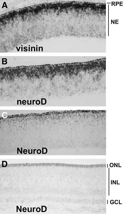
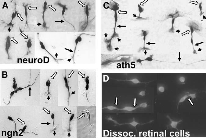
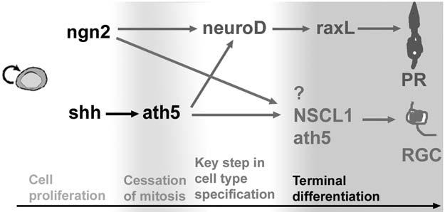
The various cell types in the vertebrate retina arise from a pool of common progenitors. The way that the cell types are specified has been a long-standing issue. Decades of research have yielded a large body of information regarding the involvement of extrinsic factors, and only recently has the function of intrinsic factors begun to emerge. This article reviews recent studies addressing the role of basic helix-loop-helix (bHLH) factors in specifying retinal cell types, with an emphasis on bHLHhierarchies leading to photoreceptor production. Photoreceptor genesis appears to employ two transcriptional pathways: ngn2-->neuroD-->raxL and ath5-->neuroD-->raxL. ngn2 and ath5 function in progenitors, which can potentially develop into different cell types. neuroD represents one of the central steps in photoreceptor specification. Ath5 is also essential for ganglion cell development. It remains to be demonstrated whether a bHLH gene functions as a key player in specifying the other types of retinal cells. Genetic knockout studies have indicated intricate cross-regulation among bHLH genes. Future studies are expected to unveil the mechanism by which bHLH factors network with intrinsic factors and communicate with extrinsic factors to ensure a balanced production of the various types of retinal cells.
Figures
Similar articles
-
A role of ath5 in inducing neuroD and the photoreceptor pathway.J Neurosci. 2004 Aug 11;24(32):7150-8. doi: 10.1523/JNEUROSCI.2266-04.2004. J Neurosci. 2004. PMID: 15306648 Free PMC article.
-
Requirement of neuroD for photoreceptor formation in the chick retina.Invest Ophthalmol Vis Sci. 2004 Jan;45(1):48-58. doi: 10.1167/iovs.03-0774. Invest Ophthalmol Vis Sci. 2004. PMID: 14691153 Free PMC article.
-
Requirement of multiple basic helix-loop-helix genes for retinal neuronal subtype specification.J Biol Chem. 2004 Jul 2;279(27):28492-8. doi: 10.1074/jbc.M400871200. Epub 2004 Apr 22. J Biol Chem. 2004. PMID: 15105417
-
Retinal cell fate determination and bHLH factors.Semin Cell Dev Biol. 2004 Feb;15(1):83-9. doi: 10.1016/j.semcdb.2003.09.005. Semin Cell Dev Biol. 2004. PMID: 15036211 Review.
-
The roles of intrinsic and extrinsic cues and bHLH genes in the determination of retinal cell fates.Curr Opin Neurobiol. 1999 Feb;9(1):37-46. doi: 10.1016/s0959-4388(99)80005-1. Curr Opin Neurobiol. 1999. PMID: 10072376 Review.
Cited by
-
Neurogenin3 promotes early retinal neurogenesis.Mol Cell Neurosci. 2009 Feb;40(2):187-98. doi: 10.1016/j.mcn.2008.10.006. Epub 2008 Nov 6. Mol Cell Neurosci. 2009. PMID: 19028584 Free PMC article.
-
Protocadherin-17 function in Zebrafish retinal development.Dev Neurobiol. 2013 Apr;73(4):259-73. doi: 10.1002/dneu.22053. Epub 2013 Jan 24. Dev Neurobiol. 2013. PMID: 22927092 Free PMC article.
-
Wnt signaling promotes regeneration in the retina of adult mammals.J Neurosci. 2007 Apr 11;27(15):4210-9. doi: 10.1523/JNEUROSCI.4193-06.2007. J Neurosci. 2007. PMID: 17428999 Free PMC article.
-
NeuroD regulates proliferation of photoreceptor progenitors in the retina of the zebrafish.Mech Dev. 2009 Mar-Apr;126(3-4):128-41. doi: 10.1016/j.mod.2008.11.009. Epub 2008 Dec 14. Mech Dev. 2009. PMID: 19121642 Free PMC article.
-
Multi-site phosphorylation regulates NeuroD4 activity during primary neurogenesis: a conserved mechanism amongst proneural proteins.Neural Dev. 2015 Jun 18;10:15. doi: 10.1186/s13064-015-0044-8. Neural Dev. 2015. PMID: 26084567 Free PMC article.
References
-
- Curcio CA. Photoreceptor topography in ageing and age-related maculopathy. Eye. 2001;15:376–383. - PubMed
-
- Turner DL, Cepko CL. A common progenitor for neurons and glial persists in rat retina late in development. Nature. 1987;328:131–136. - PubMed
-
- Raymond PA. Cell determination and positional cues in the teleost retina: development of photoreceptors and horizontal cells. In: Lam D-K, Shatz CJ, editors. Development of the Visual System. The MIT Press; Cambridge, MA: 1991. pp. 59–78.
-
- Reh TA. Determination of cell fate during retinal histogenesis: Intrinsic and extrinsic mechanisms. In: Lam D-K, Shatz CJ, editors. Development of the Visual System. The MIT Press; Cambridge, MA: 1991. pp. 79–94.
-
- Adler R, Hatlee M. Plasticity and differentiation of embryonic retinal cells after terminal mitosis. Science. 1989;243:391–393. - PubMed
Publication types
MeSH terms
Substances
Grants and funding
LinkOut - more resources
Full Text Sources
