. 2005 Oct 10;6(1):114.
doi: 10.1186/1465-9921-6-114.

Rhinovirus infection induces cytotoxicity and delays wound healing in bronchial epithelial cells

Affiliations

Rhinovirus infection induces cytotoxicity and delays wound healing in bronchial epithelial cells

Apostolos Bossios et al. Respir Res. .

Abstract

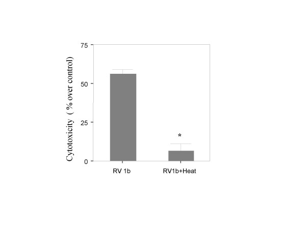
Background: Human rhinoviruses (RV), the most common triggers of acute asthma exacerbations, are considered not cytotoxic to the bronchial epithelium. Recent observations, however, have questioned this knowledge. The aim of this study was to evaluate the ability of RV to induce epithelial cytotoxicity and affect epithelial repair in-vitro.

Methods: Monolayers of BEAS-2B bronchial epithelial cells, seeded at different densities were exposed to RV serotypes 1b, 5, 7, 9, 14, 16. Cytotoxicity was assessed chromatometrically. Epithelial monolayers were mechanically wounded, exposed or not to RV and the repopulation of the damaged area was assessed by image analysis. Finally epithelial cell proliferation was assessed by quantitation of proliferating cell nuclear antigen (PCNA) by flow cytometry.

Results: RV1b, RV5, RV7, RV14 and RV16 were able to induce considerable epithelial cytotoxicity, more pronounced in less dense cultures, in a cell-density and dose-dependent manner. RV9 was not cytotoxic. Furthermore, RV infection diminished the self-repair capacity of bronchial epithelial cells and reduced cell proliferation.

Conclusion: RV-induced epithelial cytotoxicity may become considerable in already compromised epithelium, such as in the case of asthma. The RV-induced impairment on epithelial proliferation and self-repair capacity may contribute to the development of airway remodeling.


Figures

Figure 1
Figure 1
Cytotoxicity of different RV serotypes; 1b, 7, 14, 5, 16 and 9, at MOI = 1 on BEAS-2B cells, cultured until reaching different densities (100, 50, 25 and 12.5%). An inverse correlation between cell density and RV-induced cytotoxicity is observed for RV1b, RV7, RV14 and RV16, (*p < 0.05, n = 3–26, linear regression).
Figure 2
Figure 2
Cytotoxicity of RV16 (A), RV5 (B) and RV1b (C) at MOI = 5 on BEAS-2B cells, cultured until reaching different densities (100, 50, 25 and 12.5%, n = 3–8). RVs became more cytotoxic at this MOI, and density dependence appeared for RV5. Dose dependence is shown for RV1b (D) at 0.5, 1, and 5 MOI for both 100% and 50% cell densities (p = 0.000 in both cases, n = 6, ANOVA).
Figure 3
Figure 3
Cytotoxicity of active and heat inactivated RV1b (MOI = 1) on 50% confluent BEAS-2B cells. Inactivated virus is no longer cytotoxic (*p = 0.021, n = 4, Mann Whitney).
Figure 4
Figure 4
Damaged epithelium (t = 0) is suboptimally repopulated after RV-infection in comparison to control BEAS-2B cells. DAPI stained cells (A). Repopulation of damaged epithelium, expressed as unpopulated area in mm2, in RV-infected and non-infected BEAS-2B cells, immediately after damage (t = 0) and at 24, 48 and 72 hours later (B). Repopulation in infected cells is significantly reduced (*p < 0.05, **p = 0.001, n = 8, Mann Whitney).
Figure 5
Figure 5
Proliferation of BEAS-2B cells, as mean fluorescence intensity (MFI) of PCNA in RV-infected and control cells at various time points after reculture (A). The proliferation rate is significantly reduced for RV-infected cells at all time points (*p = 0.012, ** p < 0.05, n = 3–7, Mann Whitney). Representative histograms at t = 0 and 24 hours are shown (B). Closed line: isotype control, thick line: RV-infected, dashed line: non-infected control cells.

References

    1. Adam EC, Holgate ST, Fildew CJ, Lackie PM. Role of carbohydrates in repair of human respiratory epithelium using an in vitro model. Clin Exp Allergy. 2003;33:1398–1404. doi: 10.1046/j.1365-2222.2003.01771.x. - DOI - PubMed
    1. Sacco O, Silvestri M, Sabatini F, Sale R, Defilippi AC, Rossi GA. Epithelial cells and fibroblasts: structural repair and remodelling in the airways. Paediatr Respir Rev. 2004;5(Suppl A):S35–S40. doi: 10.1016/S1526-0542(04)90008-5. - DOI - PubMed
    1. Woltmann G, Ward RJ, Symon FA, Rew DA, Pavord ID, Wardlaw AJ. Objective quantitative analysis of eosinophils and bronchial epithelial cells in induced sputum by laser scanning cytometry. Thorax. 1999;54:124–130. - PMC - PubMed
    1. Bousquet J, Jeffery PK, Busse WW, Johnson M, Vignola AM. Asthma. From bronchoconstriction to airways inflammation and remodeling. Am J Respir Crit Care Med. 2000;161(5):1720–1745. - PubMed
    1. Davies DE, Wicks J, Powell RM, Puddicombe SM, Holgate ST. Airway remodeling in asthma: new insights. J Allergy Clin Immunol. 2003;111(2):215–225. doi: 10.1067/mai.2003.128. - DOI - PubMed

Substances

LinkOut - more resources