Comparative Study
. 2005 Oct 17;202(8):1051-61.
doi: 10.1084/jem.20040662. Epub 2005 Oct 10.

Essential role for CD103 in the T cell-mediated regulation of experimental colitis

Affiliations
Comparative Study

Essential role for CD103 in the T cell-mediated regulation of experimental colitis

Oliver Annacker et al. J Exp Med. .

Abstract

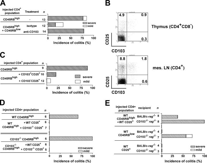
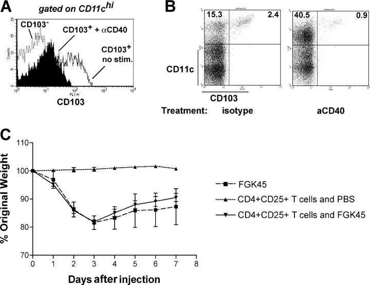
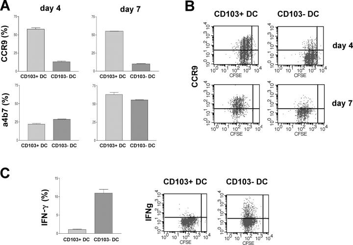
The integrin CD103 is highly expressed at mucosal sites, but its role in mucosal immune regulation remains poorly understood. We have analyzed the functional role of CD103 in intestinal immune regulation using the T cell transfer model of colitis. Our results show no mandatory role for CD103 expression on T cells for either the development or CD4+CD25+ regulatory T (T reg) cell-mediated control of colitis. However, wild-type CD4+CD25+ T cells were unable to prevent colitis in immune-deficient recipients lacking CD103, demonstrating a nonredundant functional role for CD103 on host cells in T reg cell-mediated intestinal immune regulation. Non-T cell expression of CD103 is restricted primarily to CD11c(high)MHC class II(high) dendritic cells (DCs). This DC population is present at a high frequency in the gut-associated lymphoid tissue and appears to mediate a distinct functional role. Thus, CD103+ DCs, but not their CD103- counterparts, promoted expression of the gut-homing receptor CCR9 on T cells. Conversely, CD103- DCs promoted the differentiation of IFN-gamma-producing T cells. Collectively, these data suggest that CD103+ and CD103- DCs represent functionally distinct subsets and that CD103 expression on DCs influences the balance between effector and regulatory T cell activity in the intestine.


Figures

Figure 1.
Figure 1.
Histopathological definition of colitis as applied throughout this study. Animals were transferred with CD4 T cell subsets and killed after 8–12 wk or when reaching 80% of their initial weight. Representative photomicrographs show hematoxylin and eosin–stained sections of colons from C.B.-17-SCID recipients of CD4 T cell subsets and represent different definitions of colitis (none, mild, and severe). Bar, 100 μm.
Figure 2.
Figure 2.
CD103 is not required for the development of colitis. C.B.-17-SCID mice received 4 × 10⁵ sorted CD4+CD45RBhigh T cells. At death, hosts were analyzed for various parameters. (A) T cell numbers recovered from various organs. Symbols represent individual mice. Data are pooled from three independent experiments. The horizontal lines represent the mean. (B) Intracytoplasmic cytokine production by T cells from the LP after overnight stimulation with anti-CD3 antibodies. The numbers shown indicate the percentage of cells in that gate. Data are representative of three individual animals per group. (C) Cumulative colitis incidence in various recipients of CD103−/− CD4+CD45RBhigh T cells. Data are pooled from seven independent experiments.
Figure 3.
Figure 3.
Protection from colitis requires expression of CD103 by host cells. Mice received 4 × 10⁵ CD4+CD45RBhigh T cells with 1 or 2 × 10⁵ CD4+CD45RBlow or CD4+CD25+ T cells. (A) Colitis incidence in C.B.-17-SCID recipients of the indicated CD4 T cell populations. Antibodies were given i.p. twice weekly for 8 wk (0.5mg/injection). Data are pooled from four independent experiments. (B) Representative CD103 expression by CD4 T cells from WT animals (n = 8). The numbers shown indicate the percentage of cells in that gate. (C) Colitis incidence in C57BL/6-RAG−/− recipients of CD4+CD45RBhigh T cells and sorted CD103+ and CD103CD4+CD25+ cell subsets. Data are pooled from two independent experiments. (D) Cumulative colitis incidence in C.B.-17-SCID recipients after transfer of the indicated CD4 T cell subsets. Data are pooled from two independent experiments. (E) Colitis development in RAG−/− recipients of WT CD4 T cell subsets as indicated. Data are pooled from five independent experiments.
Figure 4.
Figure 4.
Phenotypic characteristics of CD11chighCD103+ cells. Spleens, MLN, or colons from BALB/c-RAG−/− or WT animals were digested with collagenase before the distribution of cell surface molecules was investigated. (A) Representative staining of CD11c and CD103 on splenic cell preparations from nonreconstituted Balb/c Rag−/− animals (n = 4) showing that the majority of CD103+ cells are CD11chigh. The majority of CD103+ cells in the MLN and colon were also CD11chigh. (B) Representative staining of various cell surface molecules on splenic preparations from nonreconstituted BALB/c-RAG−/− animals (n = 4). FSC, forward scatter. (C) Representative staining of CD11b, CD8α, and CD103 on MLN and colon preparations from nonreconstituted Balb/c Rag−/− animals (n = 6, pooled into three groups). Positioning of quadrants reflects isotype controls. The numbers shown in A–C indicate the percentage of cells in that gate. (D) Percentage of CD11chigh cells in the spleen (n = 7), MLN (n = 10, pooled into five groups), and colon (n = 10, pooled into five groups) expressing CD103, as determined by FACS analysis. Values represent mean ± SD.
Figure 5.
Figure 5.
Effect of stimulation through CD40 on CD103 expression. (A) FACS-sorted CD11chigh subpopulations were incubated overnight with 5 μg/ml anti-CD40 mAbs before assessing the expression of CD103. Representative stainings of two independent experiments per group. Further experiments with less stringently sorted CD11chigh populations gave similar results. (B) C57BL/6-RAG−/− mice were injected i.p. with 200 μg anti-CD40 or isotype control mAbs. 3 d later, spleens were analyzed for the presence of CD103+ and CD11c+ cells. The numbers shown indicate the percentage of cells in that gate. Stainings are representative of three animals per group. (C) Wasting disease in mice treated with anti-CD40 in the presence or absence of CD4+CD25+ cells. Wasting was accompanied by colitis. Transfer of CD4+CD25+ T cells 28 d before injection of anti-CD40 did not result in any improvement in wasting or in the severity of colitis. Data is representative of three similar experiments. Values represent mean ± SD.
Figure 6.
Figure 6.
Functional characteristics of CD11chigh CD103+ cells. MACS-sorted, CFSE-labeled CD4+ cells from DO11.10 SCID donors were incubated with FACS-sorted CD103+CD11chigh or CD103CD11chigh DCs from MLN of Balb/c donors and OVA peptide. T cells were analyzed by FACS for expression of CCR9 and α4β7 at day 4 of culture and, after a further 3 d of expansion in IL-2. At day 7 of culture, T cells were restimulated overnight with platebound anti-CD3 mAb before intracellular cytokines were assessed. (A) Mean percentage ± SD of T cells expressing CCR9 and α4β7. Data from one of two independent experiments. (B) Representative staining of CCR9 on T cells. (C) Mean percentage ± SD of T cells producing IFN-γ and representative stainings. Data from one of two independent experiments.

References

    1. Morrissey, P.J., K. Charrier, S. Braddy, D. Liggitt, and J.D. Watson. 1993. CD4+ T cells that express high levels of CD45RB induce wasting disease when transferred into congenic severe combined immunodeficient mice. Disease development is prevented by cotransfer of purified CD4+ T cells. J. Exp. Med. 178:237–244. - PMC - PubMed
    1. Powrie, F., M.W. Leach, S. Mauze, L.B. Caddle, and R.L. Coffman. 1993. Phenotypically distinct subsets of CD4+ T cells induce or protect from chronic intestinal inflammation in C. B-17 scid mice. Int. Immunol. 5:1461–1471. - PubMed
    1. Singh, B., S. Read, C. Asseman, V. Malmstrom, C. Mottet, L.A. Stephens, R. Stepankova, H. Tlaskalova, and F. Powrie. 2001. Control of intestinal inflammation by regulatory T cells. Immunol. Rev. 182:190–200. - PubMed
    1. Sakaguchi, S., N. Sakaguchi, J. Shimizu, S. Yamazaki, T. Sakihama, M. Itoh, Y. Kuniyasu, T. Nomura, M. Toda, and T. Takahashi. 2001. Immunologic tolerance maintained by CD25+ CD4+ regulatory T cells: their common role in controlling autoimmunity, tumor immunity, and transplantation tolerance. Immunol. Rev. 182:18–32. - PubMed
    1. Shevach, E.M. 2002. CD4+ CD25+ suppressor T cells: more questions than answers. Nat. Rev. Immunol. 2:389–400. - PubMed

Publication types

MeSH terms