. 2005 Oct 10;171(1):153-64.
doi: 10.1083/jcb.200506152.

Integrin-dependent actomyosin contraction regulates epithelial cell scattering

Affiliations

Integrin-dependent actomyosin contraction regulates epithelial cell scattering

Johan de Rooij et al. J Cell Biol. .

Abstract

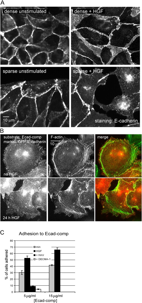
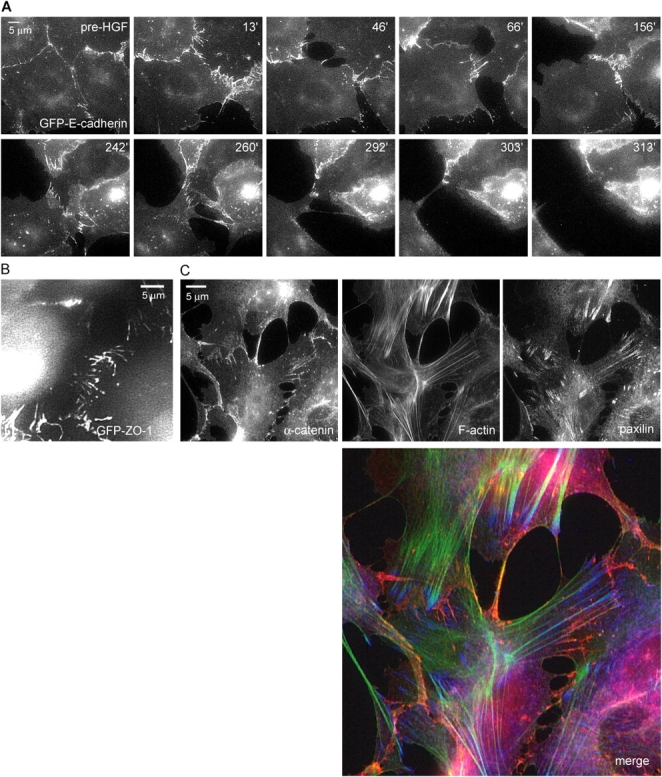
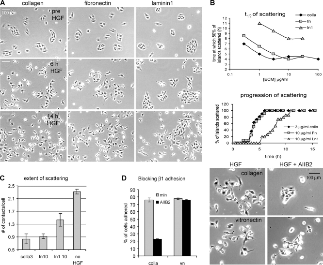
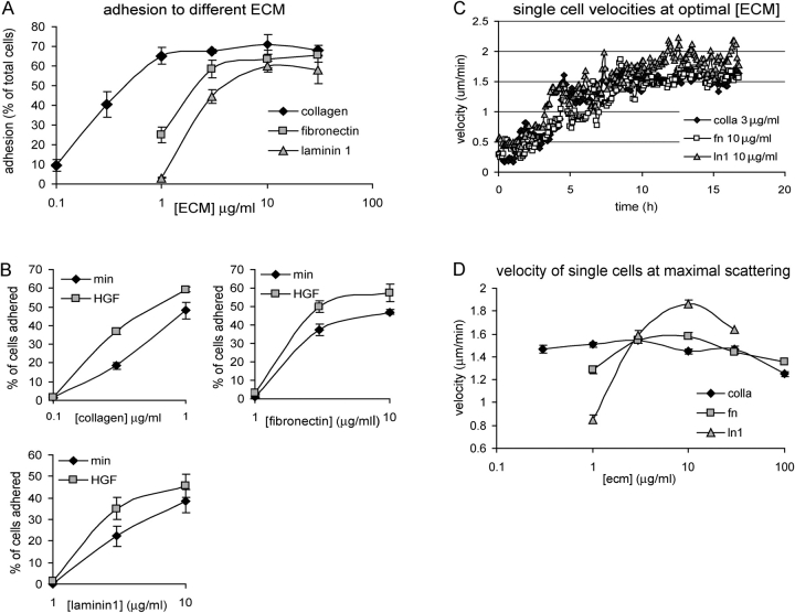
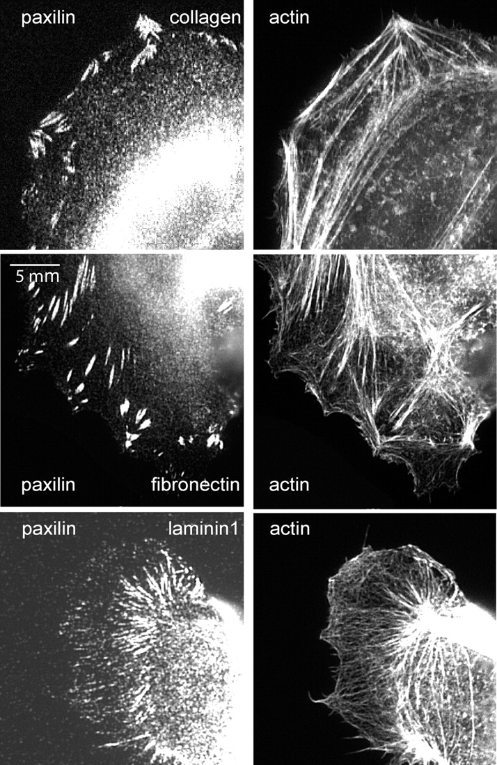
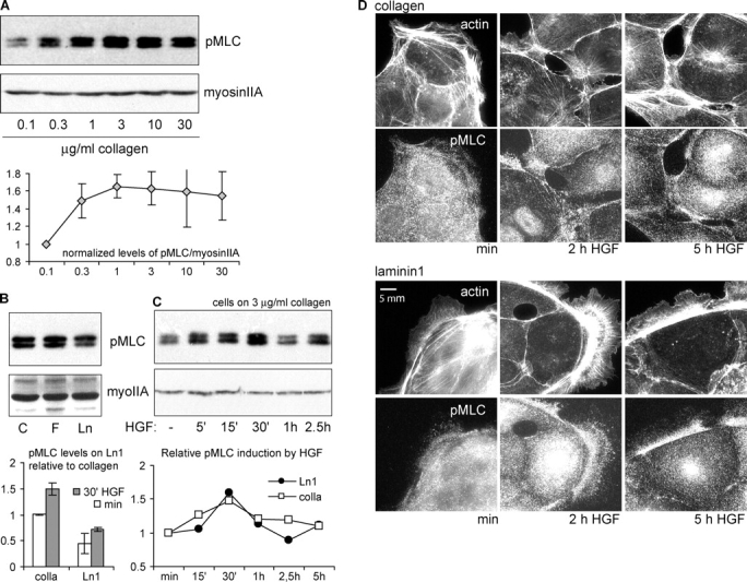
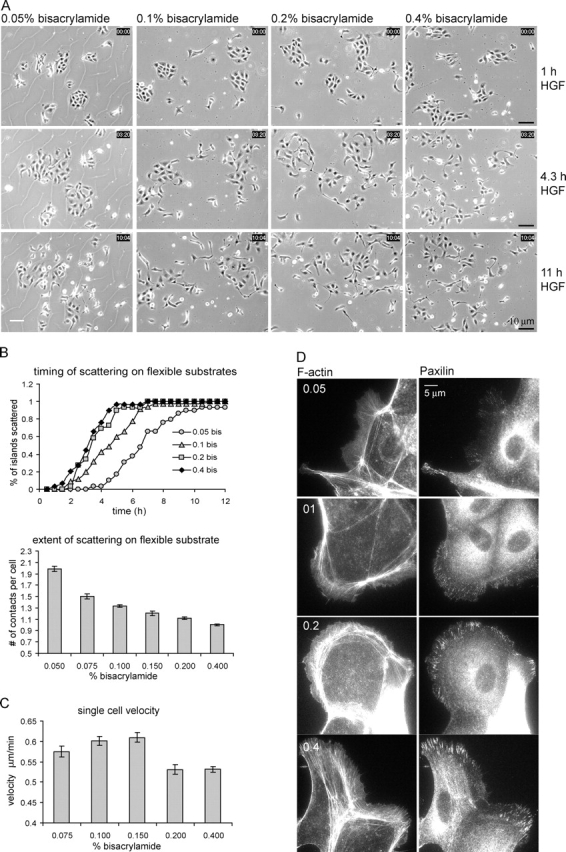
The scattering of Madin-Darby canine kidney cells in vitro mimics key aspects of epithelial-mesenchymal transitions during development, carcinoma cell invasion, and metastasis. Scattering is induced by hepatocyte growth factor (HGF) and is thought to involve disruption of cadherin-dependent cell-cell junctions. Scattering is enhanced on collagen and fibronectin, as compared with laminin1, suggesting possible cross talk between integrins and cell-cell junctions. We show that HGF does not trigger any detectable decrease in E-cadherin function, but increases integrin-mediated adhesion. Time-lapse imaging suggests that tension on cell-cell junctions may disrupt cell-cell adhesion. Varying the density and type of extracellular matrix proteins shows that scattering correlates with stronger integrin adhesion and increased phosphorylation of the myosin regulatory light chain. To directly test the role of integrin-dependent traction forces, substrate compliance was varied. Rigid substrates that produce high traction forces promoted scattering, in comparison to more compliant substrates. We conclude that integrin-dependent actomyosin traction force mediates the disruption of cell-cell adhesion during epithelial cell scattering.


Figures

Figure 1.
Figure 1.
HGF does not inhibit the ability of E-cadherin to form homotypic interactions. (A) Lack of effect of HGF on E-cadherin in cell–cell junctions. MDCK cells plated at high or low density on Cn-coated coverslips were treated with HGF for 24 h. Cells were fixed and E-cadherin was immunolocalized. (B) Lack of effect of HGF on GFP-E-cadherin distribution on E-cadherin–coated coverslips. GFP-E-cadherin–expressing MDCK cells were plated on Ecad-comp in the absence of HGF for 3 h or in the presence of HGF for 24 h, fixed, and stained for F-actin. (C) HGF increases adhesion to Ecad-comp. MDCK cells were plated on the indicated amounts of Ecad-comp in the absence or presence of HGF or inhibitors (DECMA-1 or HAV-peptide) and treated and quantified as described in Materials and methods. Data are means ± SD; n = 3.
Figure 2.
Figure 2.
Cell–cell adhesions are pulled apart during scattering. (A) Visualizing GFP-cadherin during scattering. MDCK cells stably expressing GFP-E-cadherin on 3 μg/ml Cn were imaged by time-lapse epifluorescence microscopy. HGF was added after 2 h and imaging continued. Images are from time-lapse Video 2. Images show dynamic adherens junctions that reorganized into radial streaks before breakage. (B) Scattering MDCK cells expressing GFP–ZO-1 were observed in the same manner as in A (Video 3). Cells just before the moment of cell–cell disruption are shown, demonstrating similar radial streaks as seen in GFP-E-cadherin. (C) Cells treated with HGF for 5 h were fixed and stained for α-catenin (red), paxillin (blue), and F-actin (green), to simultaneously visualize adherens junction, focal adhesions, and the actin cytoskeleton. In cells with disrupting adherens junctions, F-actin bundles terminate in focal adhesions just adjacent to cell–cell junctions.
Figure 3.
Figure 3.
Scattering is promoted by increasing ECM concentration and is more efficient on Cn and Fn than on Ln 1. MDCK cells plated on the indicated ECM proteins were imaged by time-lapse phase-contrast microscopy. HGF was added after 2 h and imaging continued for 16 h. (A) Representative images from the time-lapse series (Video 4) showing the differences between scattering on the three different ECM proteins at saturating concentrations (3 μg/ml type I Cn, 10 μg/ml Fn, 10 μg/ml Ln1). By 6 h, cells have begun to scatter on Cn and Fn, but have not initiated on Ln1. (B) For all matrices, t 1/2 of scattering is reached faster on increasing matrix concentration and is saturable. Quantification of scattering from three time-lapses per condition from one representative experiment is shown. The top graph shows the time at which 50% of the islands initiated scattering (as measured by the disruption of at least three cell–cell junctions per island). The bottom graph depicts the progression of scattering at saturating ECM concentrations. Scattering progresses with similar kinetics on Fn and Cn, and much more slowly on Ln1. (C) Cells on Ln 1 do not scatter as completely as cells on other matrices. The extent of scattering, quantified as the average number of cell–cell contacts per cell at 14 h of HGF on the indicated matrix (when scattering was complete) or in the absence of HGF on 3 μg/ml Cn. Data are means ± SEM, and at least 250 cells were counted per condition. (D) Scattering is not specific to β1 integrins. Cells were induced to scatter as in A on 3 μg/ml Cn or 3 μg/ml Vn in the absence or presence of the β1 integrin–blocking antibody AIIB2 (10 μg/ml) and followed by time-lapse imaging. Three time-lapse series with identical results were obtained (Video 5) and representative images at 12 h after HGF are shown (right). As a control, the effect of AIIB2 on the adhesion to Cn and Vn was measured using an adhesion assay (left), showing that β1 integrins are not involved in the adhesion to Vn. Data are means ± SD; n = 3.
Figure 4.
Figure 4.
Scattering on increasing ECM correlates with adhesion strength, not migration velocity. (A and B) MDCK cells plated on different amounts of ECM, as in Fig. 3, were allowed to adhere in the presence or absence of HGF for 1 h. Unbound cells were removed and bound cells were quantified. Data are means ± SD; n = 3. Results show that adhesion on all matrices is saturable, is in the order Cn > Fn > Ln1, and is increased by HGF. (C) The average velocity of single cells on saturating concentrations of each ECM was determined from the same data used for Fig. 3 B. For all matrices, HGF induces an increase in cell migration, similar in timing and magnitude. (D) Cell velocity exhibits a classic biphasic response to increasing concentrations of all matrices. The average single cell velocity between 12–16 h after HGF was calculated for increasing ECM concentrations. At this interval, velocity had reached its maximum in all cases. Data are means ± SEM; at least 30 single cells per condition were included in this measurement.
Figure 5.
Figure 5.
Focal adhesions and F-actin on different types of ECM. Paxillin and F-actin localization in cells grown on 3 μg/ml Cn, 10 μg/ml Fn, or 10 μg/ml Ln1. In cells on Fn and Cn, larger peripheral focal adhesions are associated with dense actin bundles. On Ln1, peripheral focal adhesions and actin bundles are nearly absent, although elongated central adhesions are associated with actin bundles.
Figure 6.
Figure 6.
Myosin II regulatory light chain phosphorylation and distribution are regulated by ECM type and concentration. (A) MLC phosphorylation increases with concentration of matrix. Lysates from cells plated on the indicated ECM were analyzed for pMLC by Western blotting. Total myosin IIA heavy chain was analyzed as a loading control. The induction of pMLC by increasing Cn was quantified relative to the lowest concentration used. Data are means ± SD; n = 3. (B) pMLC is greater in cells plated on Cn or Fn than on Ln 1. pMLC content was compared in cells on saturating concentrations of 3 μg/ml Cn, 10 μg/ml Fn, and 10 μg/ml Ln1. Levels of pMLC on Ln1 relative to Cn in the absence and presence of HGF (30 min) were calculated. Data are means ± SD; n = 5. (C) HGF induces a transient increase in pMLC that peaks at 30 min after application. Cells on Cn (3 μg/ml) or Ln1 (10 μg/ml) were stimulated with HGF for the indicated periods of time. Levels of pMLC relative to unstimulated samples on either Cn or Ln1 are shown. (D) HGF induces localization of pMLC to actin bundles near cell–cell junctions on Cn, but not on Ln1. Cells were plated on either 3 μg/ml Cn- or 10 μg/ml Ln1-coated coverslips and stimulated for the indicated times with HGF. Cells were fixed, and actin and pMLC were localized.
Figure 7.
Figure 7.
Substrate rigidity regulates scattering. (A–C) Cells were plated for 20 h on Fn-coated acrylamide substrates of increasing rigidity (determined by the percentage of bisacrylamide), stimulated with HGF, and observed by phase-contrast timelapse imaging. (A) Representative pictures of key time points after HGF stimulation show increased scattering on more rigid substrates (B; Video 6). (top) Scattering time course, quantified as in Fig. 3 A. (bottom) Extent of scattering at 10 h HGF, quantified as in Fig. 3 C. Data are means ± SEM. (C) Single cell migration exhibits a biphasic response to substrate stiffness. Velocity was determined at 8–10 h after HGF stimulation, when maximal velocity was reached. Data are means ± SEM. The software was unable to track cells grown at the 0.05% bisacrylamide-containing substrate because of cracks in the substrate (as in A, left) interfering in the segmentation algorithm. (D) Substrate stiffness promotes the formation of larger peripheral adhesions associated with thick actin bundles. Cells on flexible substrates for 16 h were fixed and stained as indicated.

References

    1. Adams, C.L, Y.T. Chen, S.J. Smith, and W.J. Nelson. 1998. Mechanisms of epithelial cell-cell adhesion and cell compaction revealed by high-resolution tracking of E-cadherin-green fluorescent protein. J. Cell Biol. 142:1105–1119 - PMC - PubMed
    1. Angres, B., A. Barth, and W.J. Nelson. 1996. Mechanism for transition from initial to stable cell–cell adhesion: kinetic analysis of E-cadherin–mediated adhesion using a quantitative adhesion assay. J. Cell Biol. 134:549–557. - PMC - PubMed
    1. Avizienyte, E., A.W. Wyke, R.J. Jones, G.W. McLean, M.A. Westhoff, V.G. Brunton, and M.C. Frame. 2002. Src-induced de-regulation of E-cadherin in colon cancer cells requires integrin signalling. Nat. Cell Biol. 4:632–638. - PubMed
    1. Avizienyte, E., V.J. Fincham, V.G. Brunton, and M.C. Frame. 2004. Src SH3/2 domain-mediated peripheral accumulation of Src and phospho-myosin is linked to deregulation of E-cadherin and the epithelial-mesenchymal transition. Mol. Biol. Cell. 15:2794–2803. - PMC - PubMed
    1. Balkovetz, D.F., and V. Sambandam. 1999. Dynamics of E-cadherin and gamma-catenin complexes during dedifferentiation of polarized MDCK cells. Kidney Int. 56:910–921. - PubMed

Publication types

MeSH terms