. 2005 Oct 18;102(42):15099-104.
doi: 10.1073/pnas.0504568102. Epub 2005 Oct 10.

Severe muscle disease-causing desmin mutations interfere with in vitro filament assembly at distinct stages

Affiliations

Severe muscle disease-causing desmin mutations interfere with in vitro filament assembly at distinct stages

Harald Bär et al. Proc Natl Acad Sci U S A. .

Abstract

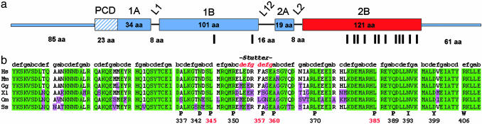
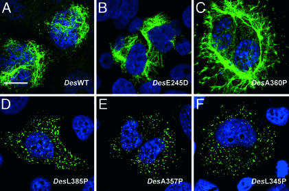
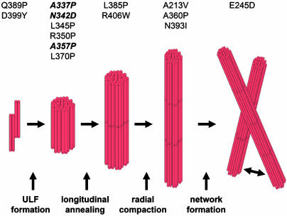
Desmin is the major intermediate filament (IF) protein of muscle. Recently, mutations of the desmin gene have been reported to cause familial or sporadic forms of human skeletal, as well as cardiac, myopathy, termed desmin-related myopathy (DRM). The impact of any of these mutations on filament assembly and integration into the cytoskeletal network of myocytes is currently not understood, despite the fact that all cause the same histopathological defect, i.e., desmin aggregation. To gain more insight into the molecular basis of this process, we investigated how mutations within the alpha-helical rod domain of desmin affect both the assembly of the recombinant protein in vitro as well as the filament-forming capacity in cDNA-transfected cells. Whereas 6 of 14 mutants assemble into seemingly normal IFs in the test tube, the other mutants interfere with the assembly process at distinct stages, i.e., tetramer formation, unit-length filament (ULF) formation, filament elongation, and IF maturation. Correspondingly, the mutants with in vitro assembly defects yield dot-like aggregates in transfected cells, whereas the mutants that form IFs constitute a seemingly normal IF cytoskeleton in the cellular context. At present, it is entirely unclear why the latter mutant proteins also lead to aggregate formation in myocytes. Hence, these findings may be a starting point to dissect the contribution of the individual subdomains for desmin pathology and, eventually, the development of therapeutic interventions.


Figures

Fig. 1.
Fig. 1.
Desmin mutations found in the α-helical rod of human desmin. (a) Schematic view of the organization of the human desmin molecule. The α-helical central rod domain is interrupted by the three non-α-helical linker regions L1, L12, and L2, which results in the formation of four α-helical segments, termed coils 1A, 1B, 2A, and 2B. The N-terminal “head” and C-terminal “tail” segments are non-α-helical. PCD, precoiled-coil domain; red, region of the molecule where most mutations are located, i.e., coil 2B. Vertical bars depict the localization of all mutations investigated. (b) Comparison of the amino acid sequence of coil 2B of various desmin proteins (Hs, Homo sapiens; Mm, Mus musculus; Gg, Gallus gallus; XL, Xenopus laevis; Om, Oncorhynchus mykiss; Ss, Scyliorhinus stellaris; the alignment was performed by using clustal). Missense mutations of desmin are depicted in the bottom line. Green, all amino acids of a column are identical; purple, amino acid in one column differs from the corresponding amino acid in Hs. Note that the sequence of murine desmin is virtually identical to the human sequence (>99%), and that most mutations reside in highly conserved regions of the last two-thirds of coil 2B. Data for mutations highlighted red are presented in Fig. 2.
Fig. 2.
Fig. 2.
Assembly properties of various desmin mutants. Electron microscopic analysis of negatively stained structures obtained from WT desmin and from representative desmin mutants after assembly in 50 mM NaCl/25 mM Tris·HCl (pH 7.5) for 10 sec, 5 min, and 60 min as indicated in comparison to viscometric profiles of assembly (Right). (Scale bar: 100 nm.) Viscosity changes were measured after 1 min and then at 5-min intervals for 60 min. Abscissa, time (minutes); ordinate, relative viscosity. The profile obtained for DesWT (▪) depicts the normal increase of relative viscosity after addition of filament forming buffer. ▵, the profiles obtained for corresponding mutant proteins. The protein concentration was 0.3 mg/ml except for DesA360P, which measured 0.1 mg/ml.
Fig. 3.
Fig. 3.
Forced expression of desmin mutants in human cultured cells. Indirect immunofluorescence microscopy of human desmin- and vimentin-free SW13 cells cDNA-transfected with WT desmin (A), DesE245D (B), DesA360P (C), DesL385P (D), DesA357P (E), and DesL345P (F). Green, immunostaining; blue, DAPI staining. Note that DesE245D and DesA360P form an apparently normal IF network in the transfected cells shown, whereas all other mutations, which exhibit a compromised in vitro filament assembly, only show intracytoplasmic aggregates. (Scale bar: 10 μm.)
Fig. 4.
Fig. 4.
Hypothetical scheme of the in vitro assembly of desmin. Mutations are indicated at the stage when first divergences from the normal assembly path become apparent. This scheme provides further support for the occurrence of distinct assembly intermediates as proposed by this model. DesQ389P and DesD399Y associate already under nonassembly conditions to complexes with sedimentation coefficients, s, higher than the normal 5.2 S. Nonetheless, they still assemble into elongated filamentous structures. Mutations depicted in bold form filamentous structures during the early phase of assembly but tend to aggregate at later time points.

References

    1. Herrmann, H. & Aebi, U. (2004) Annu. Rev. Biochem. 73, 749–789. - PubMed
    1. Lazarides, E. & Hubbard, B. D. (1976) Proc. Natl. Acad. Sci. USA 73, 4344–4348. - PMC - PubMed
    1. Bär, H., Strelkov, S. V., Sjoberg, G., Aebi, U. & Herrmann, H. (2004) J. Struct. Biol. 148, 137–152. - PubMed
    1. Bennardini, F., Wrzosek, A. & Chiesi, M. (1992) Circ. Res. 71, 288–294. - PubMed
    1. Omary, M. B., Coulombe, P. A. & McLean, W. H. (2004) N. Engl. J. Med. 351, 2087–2100. - PubMed

Publication types

MeSH terms