. 2005 Oct 18;102(42):15030-5.
doi: 10.1073/pnas.0507533102. Epub 2005 Oct 10.

RGS4 and RGS5 are in vivo substrates of the N-end rule pathway

Affiliations

RGS4 and RGS5 are in vivo substrates of the N-end rule pathway

Min Jae Lee et al. Proc Natl Acad Sci U S A. .

Abstract

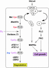
The ATE1-encoded Arg-transferase mediates conjugation of Arg to N-terminal Asp, Glu, and Cys of certain eukaryotic proteins, yielding N-terminal Arg that can act as a degradation signal for the ubiquitin-dependent N-end rule pathway. We have previously shown that mouse ATE1-/- embryos die with defects in heart development and angiogenesis. Here, we report that the ATE1 Arg-transferase mediates the in vivo degradation of RGS4 and RGS5, which are negative regulators of specific G proteins whose functions include cardiac growth and angiogenesis. The proteolysis of these regulators of G protein signaling (RGS) proteins was perturbed either by hypoxia or in cells lacking ubiquitin ligases UBR1 and/or UBR2. Mutant RGS proteins in which the conserved Cys-2 residue could not become N-terminal were long-lived in vivo. We propose a model in which the sequential modifications of RGS4, RGS5, and RGS16 (N-terminal exposure of their Cys-2, its oxidation, and subsequent arginylation) act as a licensing mechanism in response to extracellular and intracellular signals before the targeting for proteolysis by UBR1 and UBR2. We also show that ATE1-/- embryos are impaired in the activation of extracellular signal-regulated kinase mitogen-activated protein kinases and in the expression of G protein-induced downstream effectors such as Jun, cyclin D1, and beta-myosin heavy chain. These results establish RGS4 and RGS5 as in vivo substrates of the mammalian N-end rule pathway and also suggest that the O2-ATE1-UBR1/UBR2 proteolytic circuit plays a role in RGS-regulated G protein signaling in the cardiovascular system.


Figures
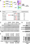
Fig. 1.
Fig. 1.
In vitro proteolysis of RGS4, RGS5, and RGS16. (A) The organization of mammalian N-end rule pathway, established from previous and current studies. N-terminal Asn and Gln function as pre-N-degrons through their deamidation by NTAN1 and a hypothetical enzyme, NTAQ1, respectively, into Asp and Glu (23). N-terminal Cys also serves as a pre-N-degron through its oxidation into Cys sulfinic acid (CysO2) or cysteic acid (CysO3) (ref. and this study). N-terminal Asp, Glu, and (oxidized) Cys function as pre-N-degrons through their N-terminal arginylation by ATE1-encoded Arg-transferase (2). The resulting N-terminal Arg together with other type 1 and type 2 N-degrons are recognized by specific E3s for subsequent Ub-dependent proteolysis. We have previously characterized UBR1 and UBR2 as such E3s (14, 16). UBR1 and UBR2 are 200 kDa-RING finger E3s that target type 1 (Arg, Lys, and His) and type 2 (Phe, Leu, Trp, Tyr, and Ile) N-degrons (14, 16, 24). In mammals, N-terminal Ala, Ser, or Thr are classified as type 3 N-degron (16), but the E3 that recognizes type 3 N-degron remains elusive. (B) RGS constructs and their expected N-terminal residues. (CD) RGS proteins were expressed in reticulocyte lysates either in the absence or presence of 2 mM dipeptides, and their degradation was analyzed by using time-course antibiotin Western blotting. Reactions were allowed for 60 min in D. Inhibition of RGS5 proteolysis by Arg-Ala diminished after 120 min perhaps because of instability of dipeptides. (E) RGS proteins were expressed in reticulocyte lysates in the presence of MG132. After 15 min, their ubiquitylation was analyzed by using anti-Ub immunoprecipitation and subsequent antibiotin Western blotting (Upper), while their expression levels were determined by using antibiotin Western blotting (Lower).
Fig. 2.
Fig. 2.
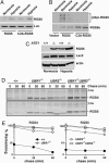
In vivo proteolysis of RGS4 and RGS5. (A) Immunoblotting of RGS4 and RGS5 transiently expressed in +/+ and ATE1–/– cells either in the absence and presence of MG132. (B) Pulse–chase analysis of RGS5, C2A-RGS5, Myc-RGS5, and RGS4. The transfected cells were labeled for 12 min with [35S]Met/[35S]Cys, followed by anti-RGS4 or RGS5 immunoprecipitation, SDS/PAGE analysis, and autoradiography. (C) Quantitation of data shown in B using PhosphorImager. (D) Immunoblotting of endogenous RGS4 in +/+ and ATE1–/– EFs. Anti-RGS5 and RGS16 antibodies did not detect endogenous proteins in either +/+ or ATE1–/– cells. (E) An ATE1 isoform and an RGS protein were coexpressed in ATE1–/– cells, followed by anti-RGS immunoblotting. A total of 4 μg of the plasmid mixture (RGS4 or RGS5, 2 μg; ATE1-1 or ATE1-2, 0, 0.25, 0.5, or 2 μg, respectively; pcDNA3, 2, 1.75, 1.5, 0 μg, respectively) was transfected into cells in a six-well plate.
Fig. 3.
Fig. 3.
Role of O2, UBR1, and UBR2 in proteolysis of RGS4 and RGS5. (A) RGS5 and C2A-RGS5 were coexpressed with luciferase in reticulocyte lysates under a normoxic or hypoxic condition, followed by anti-RGS5 or biotin-based Western blotting. (B Upper) RGS5 and C2A-RGS5 were expressed in reticulocyte lysates in the presence of MG132 either under a normoxic or hypoxic condition, followed by anti-Ub immunoprecipitation and a subsequent antibiotin Western blotting. (B) (Bottom) Comparison of RGS5 expression using anti-biotin Western blotting. (C) RGS5 was coexpressed with LacZ in +/+ and ATE1–/– EFs in normoxic and hypoxic (0.1% O2) condition, followed by anti-RGS5, LacZ, and actin immunoblotting. (D) Pulse–chase analysis of RGS4 and RGS5 in +/+, UBR1–/–, UBR2–/–, and UBR1–/–UBR2–/– cells. The transfected cells were labeled for 12 min with [35S]Met/[35S]Cys, followed by anti-RGS4 or RGS5 immunoprecipitation, SDS/PAGE analysis, and autoradiography. (E) Quantitation of data in D using PhosphorImager.
Fig. 4.
Fig. 4.
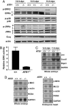
Analysis of the Gq- or Gi-activated downstream effectors in +/+ and ATE1–/– embryos. (A) The +/+ and ATE1–/– embryonic extracts were subjected to immunoblotting with antibodies against MAPKs, phospho-MAPKs, Gaq, and actin. As expected, phospho-ERK1 was not detected in mouse embryonic extracts. (B) The 12.5-dpc +/+ and ATE1–/– embryonic extracts were subjected to in vitro ERK kinase assay. (C) Total RNAs from +/+ and ATE1–/– embryos at 11.5 and 12.5 dpc were subjected to Northern blotting. (D) Total RNAs from 12.5-dpc +/+ and ATE1–/– embryos and embryonic hearts were subjected to Northern blotting.
Fig. 5.
Fig. 5.
A model for the role of the N-end rule pathway in the RGS-regulated G protein pathways.

Similar articles

Cited by

References

    1. Bachmair, A., Finley, D. & Varshavsky, A. (1986) Science 234, 179–186. - PubMed
    1. Kwon, Y. T., Kashina, A. S. & Varshavsky, A. (1999) Mol. Cell. Biol. 19, 182–193. - PMC - PubMed
    1. Kwon, Y. T., Kashina, A. S., Davydov, I. V., Hu, R. G., An, J. Y., Seo, J. W., Du, F. & Varshavsky, A. (2002) Science 297, 96–99. - PubMed
    1. Neves, S. R., Ram, P. T. & Iyengar, R. (2002) Science 296, 1636–1639. - PubMed
    1. Davydov, I. V. & Varshavsky, A. (2000) J. Biol. Chem. 275, 22931–22941. - PubMed

Publication types