Carbonic anhydrase isozyme-II-deficient mice lack the duodenal bicarbonate secretory response to prostaglandin E2
- PMID: 16217040
- PMCID: PMC1257747
- DOI: 10.1073/pnas.0508007102
Carbonic anhydrase isozyme-II-deficient mice lack the duodenal bicarbonate secretory response to prostaglandin E2
Abstract
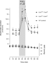
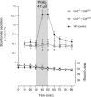
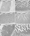
Duodenal bicarbonate secretion (DBS) is accepted as the primary mucosal defense against acid discharged from the stomach and is impaired in patients with duodenal ulcer disease. The secretory response to luminal acid is the main physiological stimulus for DBS and involves mediation by PGE2 produced by mucosal cells. The aim of this investigation is to elucidate the role of carbonic anhydrases (CAs) II and IX in PGE2-mediated bicarbonate secretion in the murine duodenum. CA II- and IX-deficient mice and different combinations of their heterozygous and WT counterparts were studied. A 10-mm segment of the proximal duodenum with intact blood supply was isolated, and DBS was titrated by pH-stat (TitroLine-easy, Schott, Mainz, Germany). Mean arterial blood pressure (MAP) was continuously recorded, and blood acid/base balance and gastrointestinal morphology were analyzed. The duodenal segment spontaneously secreted HCO3(-) at a steady basal rate of 5.3 +/- 0.6 micromol x cm(-1) x h(-1). Perfusing the duodenal lumen for 20 min with 47 microM PGE2 caused a significant increase in DBS to 13.0 +/- 2.9 micromol x cm(-1) x h(-1), P < 0.0001. The DBS response to PGE2 was completely absent in Car2-/- mice, whereas basal DBS was normal. The CA IX-deficient mice with normal Car2 alleles showed a slight increase in DBS. Histological abnormalities were observed in the gastroduodenal epithelium in both CA II- and IX-deficient mice. Our data demonstrate a gastrointestinal phenotypic abnormality associated with CA II deficiency. The results show that the stimulatory effect of the duodenal secretagogue PGE2 completely depends on CA II.
Figures


Similar articles
-
Carbonic anhydrase gene expression in CA II-deficient (Car2-/-) and CA IX-deficient (Car9-/-) mice.J Physiol. 2006 Mar 1;571(Pt 2):319-27. doi: 10.1113/jphysiol.2005.102590. Epub 2006 Jan 5. J Physiol. 2006. PMID: 16396925 Free PMC article.
-
Duodenal acidity "sensing" but not epithelial HCO3- supply is critically dependent on carbonic anhydrase II expression.Proc Natl Acad Sci U S A. 2009 Aug 4;106(31):13094-9. doi: 10.1073/pnas.0901488106. Epub 2009 Jul 21. Proc Natl Acad Sci U S A. 2009. PMID: 19622732 Free PMC article.
-
Low-dose PGE2 mimics the duodenal secretory response to luminal acid in mice.Am J Physiol Gastrointest Liver Physiol. 2004 Jun;286(6):G891-8. doi: 10.1152/ajpgi.00458.2003. Epub 2004 Feb 5. Am J Physiol Gastrointest Liver Physiol. 2004. PMID: 14764447
-
Duodenal epithelial sensing of luminal acid: role of carbonic anhydrases.Acta Physiol (Oxf). 2011 Jan;201(1):85-95. doi: 10.1111/j.1748-1716.2010.02166.x. Acta Physiol (Oxf). 2011. PMID: 20632999 Review.
-
Membrane-bound carbonic anhydrases are key pH regulators controlling tumor growth and cell migration.Adv Enzyme Regul. 2010;50(1):20-33. doi: 10.1016/j.advenzreg.2009.10.005. Epub 2009 Nov 4. Adv Enzyme Regul. 2010. PMID: 19895836 Review. No abstract available.
Cited by
-
Data Mining FAERS to Analyze Molecular Targets of Drugs Highly Associated with Stevens-Johnson Syndrome.J Med Toxicol. 2015 Jun;11(2):265-73. doi: 10.1007/s13181-015-0472-1. J Med Toxicol. 2015. PMID: 25876064 Free PMC article.
-
Carbonic Anhydrases II and IX in Non-ampullary Duodenal Adenomas and Adenocarcinoma.J Histochem Cytochem. 2021 Nov;69(11):677-690. doi: 10.1369/00221554211050133. Epub 2021 Oct 12. J Histochem Cytochem. 2021. PMID: 34636283 Free PMC article.
-
Pharmacological correction of a defect in PPAR-gamma signaling ameliorates disease severity in Cftr-deficient mice.Nat Med. 2010 Mar;16(3):313-8. doi: 10.1038/nm.2101. Epub 2010 Feb 14. Nat Med. 2010. PMID: 20154695 Free PMC article.
-
Carbonic anhydrase 2 deficiency leads to increased pyelonephritis susceptibility.Am J Physiol Renal Physiol. 2014 Oct 1;307(7):F869-80. doi: 10.1152/ajprenal.00344.2014. Epub 2014 Aug 20. Am J Physiol Renal Physiol. 2014. PMID: 25143453 Free PMC article.
-
Genetic ablation of carbonic anhydrase IX disrupts gastric barrier function via claudin-18 downregulation and acid backflux.Acta Physiol (Oxf). 2018 Apr;222(4):e12923. doi: 10.1111/apha.12923. Epub 2017 Oct 19. Acta Physiol (Oxf). 2018. PMID: 28748627 Free PMC article.
References
-
- Allen, A. & Flemström, G. (2005) Am. J. Physiol. 288, C1-C19. - PubMed
-
- Isenberg, J. I., Selling, J. A., Hogan, D. L. & Koss, M. A. (1987) N. Engl. J. Med. 316, 374-379. - PubMed
-
- Flemström, G. & Isenberg, J. I. (2001) News Physiol. Sci. 16, 23-28. - PubMed
-
- Flemström, G. & Kivilaakso, E. (1983) Gastroenterology 84, 787-794. - PubMed
-
- Flemström, G. (1994) in Physiology of the Gastrointestinal Tract, eds. Johnson, L. R., Christensen, J. J., Alpers, D. & Walsh, J. H. (Raven, New York), pp. 1285-1309.
Publication types
MeSH terms
Substances
LinkOut - more resources
Full Text Sources
Other Literature Sources
Molecular Biology Databases

