

Comparative Study
doi: 10.1212/01.wnl.0000179003.95838.71.
Three-dimensional preoperative maps of hippocampal atrophy predict surgical outcomes in temporal lobe epilepsy
Affiliations
- PMID: 16217065
- PMCID: PMC2770433
- DOI: 10.1212/01.wnl.0000179003.95838.71
Item in Clipboard
Comparative Study
Three-dimensional preoperative maps of hippocampal atrophy predict surgical outcomes in temporal lobe epilepsy
Neurology.
.
Abstract
The authors used surface-based anatomic mapping to detect features of hippocampal anatomy that correlated with surgical outcomes in patients undergoing surgery for mesial temporal lobe epilepsy with hippocampal sclerosis. Compared with a seizure-free group, hippocampal profiles for the non-seizure-free group had greater diffuse ipsilateral atrophy and more region-specific contralateral atrophy in the anterior, lateral hippocampus. These atrophic regions may indicate areas of increased epileptogenicity, contributing to poorer surgical outcomes.
Conflict of interest statement
Disclosure: The authors report no conflicts of interest.
Figures
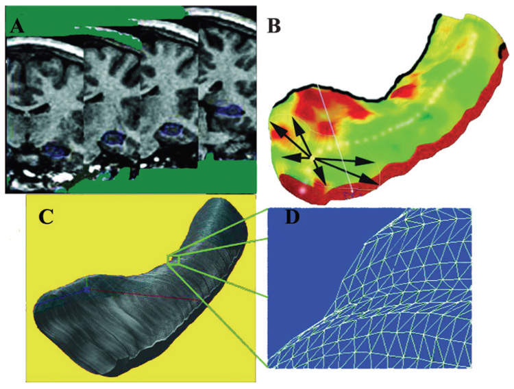
Steps involved in three-dimensional hippocampal modeling. Each individual’s hippocampus is traced in consecutive coronal MRI sections (A) and converted to a three-dimensional parametric surface (B) in which the radial size of the hippocampus is measured from a centerline and plotted in color on the surface, to index radial atrophy. These meshes are averaged across subjects (C) and atrophy relative to the contralateral hippocampus in each surgical outcome group (seizure free and not seizure free) and across the two surgical outcome groups is computed at each surface grid point (D)
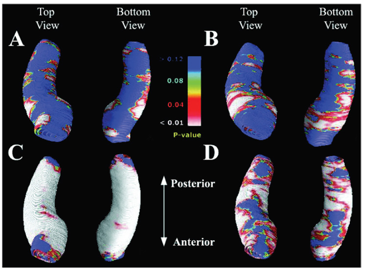
Maps identifying regions where seizure-free (SF) and non– seizure-free (NSF) surgical outcome groups differ in their degree of atrophy (A and B) and regions of hippocampal asymmetry in each surgical outcome group (C and D). Group difference maps show mean hippocampal volume differences ipsilateral (A) and contralateral (B) to the side of seizure onset. Areas of significant atrophy between the two surgical outcome groups are plotted as a map of p values. The NSF groups show significantly greater diffuse atrophy in the ipsilateral hippocampus (A), whereas the contralateral side shows more region-specific atrophy pattern (B). Maximal atrophy is seen in the anterior and lateral aspects of the contralateral hippocampus. The average distribution of atrophy was computed for patients with seizure-free postsurgical outcome (C) and those who continued to have seizures (D) by directly comparing the ipsilateral to the contralateral hippocampus. Areas of significant asymmetry are visualized as a map of p values. Both SF and NSF groups showed severe diffuse deficits along the entire hippocampus. The overall asymmetry pattern suggests that the NSF group (D) had a lesser degree of asymmetry when compared with the SF group (C). However, the anterior to posterior distribution of the asymmetry pattern does not differentiate the two surgical groups.
Comment in
-
Higher resolution MRI and image modeling for predicting surgical outcome in partial epilepsy.Neurology. 2005 Oct 11;65(7):975. doi: 10.1212/01.wnl.0000183697.64891.b4. Neurology. 2005. PMID: 16220587 No abstract available.
References
-
- Wiebe S, Blume WT, Girvin JP, Eliasziw M. A randomized, controlled trial of surgery for temporal-lobe epilepsy. N Engl J Med. 2001;345:311–318. - PubMed
-
- Hardy SG, Miller JW, Holmes MD, et al. Factors predicting outcome of surgery for intractable epilepsy with pathologically verified mesial temporal sclerosis. Epilepsia. 2003;44:565–568. - PubMed
-
- Quigg M, Bertram EH, Jackson T, Laws E. Volumetric magnetic resonance imaging evidence of bilateral hippocampal atrophy in mesial temporal lobe epilepsy. Epilepsia. 1997;38:588–594. - PubMed
-
- Spencer DD, Spencer SS, Mattson RH, et al. Access to the posterior medial temporal lobe structures in the surgical treatment of temporal lobe epilepsy. Neurosurgery. 1984;15:667–671. - PubMed
Publication types
MeSH terms
Grants and funding
LinkOut - more resources
Full Text Sources
Medical