. 2006 Jan 15;107(2):575-83.
doi: 10.1182/blood-2004-11-4377. Epub 2005 Oct 11.

Regulation of LIM-kinase 1 and cofilin in thrombin-stimulated platelets

Affiliations

Regulation of LIM-kinase 1 and cofilin in thrombin-stimulated platelets

Dharmendra Pandey et al. Blood. .

Abstract

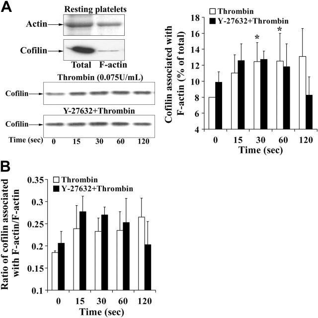
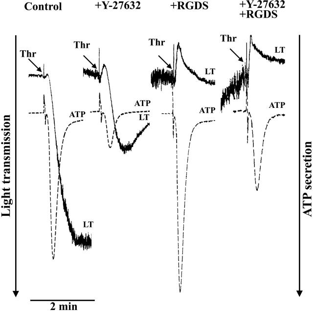
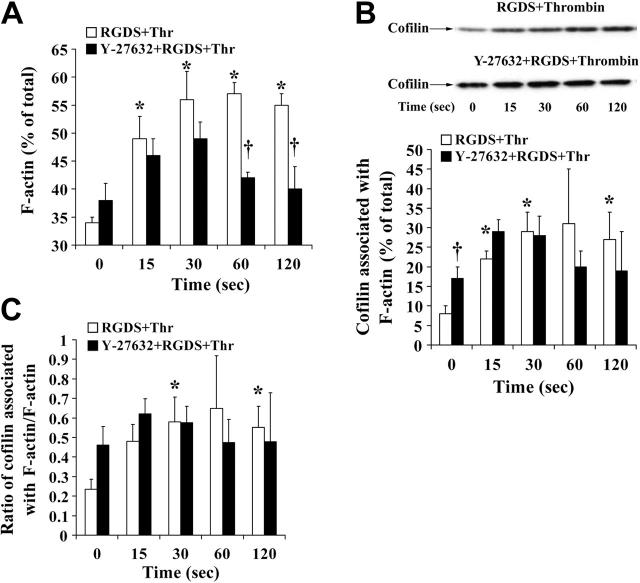
Cofilin is a regulator of actin filament dynamics. We studied whether during platelet activation Rho kinase stimulates LIM kinase (LIMK) leading to subsequent phosphorylation and inactivation of cofilin. Platelet shape change and aggregation/secretion were induced by low and high concentrations of thrombin, respectively. We found that during these platelet responses Rho kinase activation was responsible for mediating rapid Thr508 phosphorylation and activation of LIMK-1 and for the F-actin increase during shape change and, in part, during secretion. Surprisingly, during shape change cofilin phosphorylation was unaltered, and during aggregation/secretion cofilin was first rapidly dephosphorylated by an okadaic acid-insensitive phosphatase and then slowly rephosphorylated by LIMK-1. LIMK-1 phosphorylation and cofilin dephosphorylation and rephosphorylation during aggregation were independent of integrin alpha(IIb)beta(3) engagement. Cofilin phosphorylation did not regulate cofilin association with F-actin and was unrelated to the F-actin increase in thrombin-activated platelets. Our study identifies LIMK-1 as being activated by Rho kinase in thrombin-stimulated platelets. Two counteracting pathways, a cofilin phosphatase and LIMK-1, are activated during platelet aggregation/secretion regulating cofilin phosphorylation sequentially and independently of integrin alpha(IIb)beta(3) engagement. Rho kinase-mediated F-actin increase during platelet shape change and secretion involves a mechanism other than LIMK-1-mediated cofilin phosphorylation, raising the possibility of another LIMK substrate regulating platelet actin assembly.


Figures

Figure 1.
Figure 1.
The Rho kinase inhibitor Y-27632 inhibits shape change, F-actin increase, and MYPT phosphorylation induced by thrombin. Platelets were incubated with Y-27632 (20 μM) or solvent (H2O) for 30 minutes and then stimulated by thrombin (0.075 U/mL). (A) Inhibition of shape change by Y-27632. Shape change was measured by the decrease in light transmission, (left) and by confocal fluorescence microscopy of platelets stained for F-actin with Alexa Fluor 546 phalloidin (right). Bar, 10 μm. (B) Effect of Y-27632 on F-actin content during shape change. Values are mean + SD of 4 independent experiments. *Statistically significant; P < .05 with respect to time (0 seconds). †Significance between control (□) and Y-27632 (▪)–treated unstimulated platelets. (C, left) Platelet lysates were immunoblotted with anti–phospho-Thr853-MYPT antibody. Graphic representation of the result of MYPT phosphorylation in thrombin (0.075 U/mL)–stimulated platelets, evaluated by densitometry. Values are mean + SD of 3 experiments with platelets from different donors. (Right) Representative Western blot of MYPT phosphorylation in the absence and presence of Y-27632.
Figure 2.
Figure 2.
Rho kinase–dependent activation of LIMK-1 without stimulation of cofilin phosphorylation during shape change. (A, top) LIMK-1 but not LIMK-2 is expressed in platelets. Resting platelets, platelets stimulated for 30 and 60 seconds with thrombin, and endothelial-cell (EC) lysates were immunoblotted with specific anti–LIMK-1 and anti–LIMK-2 antibodies. (Bottom) Identification of cofilin in its unphosphorylated and phosphorylated states in resting platelets. Resting platelet lysates were subjected to isoelectric focusing (IEF) electrophoresis and subsequently immunoblotted with anti-cofilin antibody. Top band is the more basic, dephosphorylated form of cofilin and bottom band is the more acidic, phosphorylated form of cofilin. (B) LIMK-1 and cofilin phosphorylation during platelet shape change induced by thrombin (0.075 U/mL). Graphic representation of the result for LIMK-1 and cofilin phosphorylation. Values are the mean + SD for 3 independent experiments. (C) Effect of Y-27632 (20 μM) on LIMK-1 phosphorylation and cofilin phosphorylation in resting platelets and during thrombin (0.075 U/mL)–induced shape change. (Left) Representative immunoblots of platelets blotted with anti–phospho-LIMK-1/LIMK-2 (Thr508/505), anti–LIMK-1, anti–P-cofilin (Ser3) and anti-cofilin antibodies. (Right) Bar diagram showing cofilin phosphorylation of nontreated (□) and Y-27632-treated (▪) platelets in control and after thrombin stimulation (120 seconds). Values for cofilin phosphorylation in resting platelets and activated platelets are mean + SD of 8 and 4 independent experiments, respectively. *Statistically significant; P < .05 with respect to nontreated control.
Figure 3.
Figure 3.
Cofilin association with F-actin during platelet shape change. (A, left, top) Representative gel and immunoblot of actin and cofilin, respectively. Total indicates whole platelets; F-actin, F-actin fraction of the same number of platelets. (Left, bottom) Immunoblot of cofilin associated with F-actin during thrombin-induced shape change. Effect of Y-27632. (Right) Graphic representation of the results. Values are the mean + SD for 4 independent experiments. *Statistically significant; P < .05 with respect to time (0 seconds) in nontreated samples. (B) Bar diagram showing the ratio of cofilin associated with F-actin to F-actin in nontreated platelets (□) and platelets treated with Y-27632 (▪) during thrombin-induced shape change. Values are the mean + SD for 4 independent experiments.
Figure 4.
Figure 4.
Inhibition of thrombin-induced aggregation and secretion by the Rho kinase inhibitor Y-27632. Platelets were stimulated with thrombin (0.5 U/mL) in the absence or presence of the integrin αIIbβ3 blocker RGDS (0.5 mM) and the Rho kinase inhibitor Y-27632 (20 μM). Representative tracings for change in light transmission (LT) and ATP secretion are shown.
Figure 5.
Figure 5.
Thrombin-induced reversible phosphorylation of MYPT, irreversible phosphorylation of LIMK-1, but reversible dephosphorylation of cofilin during platelet aggregation and secretion: LIMK-1 phosphorylation and changes of cofilin phosphorylation are independent of integrin αIIbβ3 engagement. Platelet suspensions were not treated (stirring [▪] and nonstirring [▵] conditions) or treated with RGDS (0.5 mM; □) for 2 minutes before stimulation with thrombin (0.5 U/mL). (A) Thrombin-induced MYPT phosphorylation. Platelet lysates were immunoblotted with anti–phospho-Thr696-MYPT antibody. (Left) Graphic representation of results. (Right) Representative immunoblot of MYPT phosphorylation. (B) Thrombin-induced LIMK-1 phosphorylation. Platelet lysates were immunoblotted with anti–phospho-LIMK-1/LIMK-2 (Thr508/505) antibody. (Left) Graphic representation. (Right) Representative immunoblots. (C) Cofilin dephosphorylation and rephosphorylation during thrombin-induced secretion/aggregation. Platelet lysates were immunoblotted with anti–phospho-cofilin antibody. (Left) Graphic representation of results. (Right) Representative immunoblots for cofilin phosphorylation. Values are mean + SD or mean – SD of 3 independent experiments.
Figure 6.
Figure 6.
Inhibition of MYPT phosphorylation, LIMK-1 phosphorylation, LIMK-1 activity, and cofilin rephosphorylation by the Rho kinase inhibitor Y-27632 during thrombin-stimulated aggregation/secretion. Platelet samples stimulated by thrombin (0.5 U/mL) in the absence or presence of the Rho kinase inhibitor Y-27632 (20 μM) were immunoblotted with anti–phospho-MYPT, anti–phospho-LIMK-1/LIMK-2 (Thr508/505), and anti–phospho-cofilin antibodies. (A) Representative immunoblots showing concomitant inhibition of MYPT and LIMK-1 phosphorylation by Y-27632. (B) Immunoblot and graphic representation of the results of cofilin phosphorylation after thrombin stimulation of platelets in the absence (□) or presence of Y-27632 (▪). Values are mean + SD for 3 independent experiments. (C) LIMK-1 phosphorylation parallels LIMK-1 activity in thrombin-stimulated platelets. Inhibition by Y-27632. Platelets were preincubated with Y-27632 (20 μM) or solvent for 30 minutes and stimulated with thrombin in the presence of RGDS. LIMK-1 from resting and activated platelets (120 seconds) was immunoprecipitated. (Top) LIMK-1 immunoprecipitates (LIMK-1 IP) were blotted with anti–LIMK-1 antibody and anti–phospho-LIMK-1/LIMK-2 (Thr508/505) antibody. (Bottom) Immunoprecipitates were assayed for LIMK-1 activity using His-cofilin as substrate. Cofilin phosphorylation was measured by blotting the samples with anti–phospho-cofilin antibodies.
Figure 7.
Figure 7.
F-actin increase and association of cofilin with F-actin during thrombin-induced secretion: effect of Y-27632. Platelets treated with H2O (□) or Y-27632 (▪) were stimulated with thrombin (0.5 U/mL) in the presence of the integrin αIIbβ3 blocker RGDS (0.5 mM) and lysed with 1% Triton X-100 for isolation of F-actin. F-actin and cofilin associated with F-actin (percentage of total) were measured. (A) F-actin content, bar diagram. (B) Cofilin association with F-actin. (Top) Representative immunoblots. (Bottom) bar diagram (C) Bar diagram showing the ratio of F-actin–associated cofilin to F-actin. Values are the mean + SD for 4 independent experiments. *Statistically significant; P < .05 with respect to nonstimulated samples at 0 seconds. †Significance between control (□) and Y-27632 (▪)–treated platelets.

Similar articles

Cited by

References

    1. Siess W. Molecular mechanisms of platelet activation. Physiol Rev. 1989;69: 58-178. - PubMed
    1. Fox JE. Regulation of platelet function by the cytoskeleton. Adv Exp Med Biol. 1993;344: 175-185. - PubMed
    1. Hartwig JH, Barkalow K, Azim A, Italiano J. The elegant platelet: signals controlling actin assembly. Thromb Haemost. 1999;82: 392-398. - PubMed
    1. Fox JE. Cytoskeletal proteins and platelet signaling. Thromb Haemost. 2001;86: 198-213. - PubMed
    1. Li Z, Kim ES, Bearer EL. Arp2/3 complex is required for actin polymerization during platelet shape change. Blood. 2002;99: 4466-4474. - PMC - PubMed

Publication types

MeSH terms