MR imaging with diffusion-weighted imaging in acute and chronic Wernicke encephalopathy
- PMID: 16219837
- PMCID: PMC7976162
MR imaging with diffusion-weighted imaging in acute and chronic Wernicke encephalopathy
Abstract
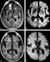
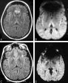
Wernicke encephalopathy is a neurologic disorder that results from thiamine deficiency. It is associated with a classic triad of symptoms consisting of ataxia, ocular motor cranial neuropathies, and changes in consciousness. We report 3 cases of Wernicke encephalopathy in which MR imaging, including diffusion-weighted imaging, was performed at the onset and during follow-up. MR imaging findings were correlated with the clinical status of both the acute and chronic stage of Wernicke encephalopathy.
Figures


Similar articles
-
MR Imaging of nonalcoholic Wernicke encephalopathy: a follow-up study.AJNR Am J Neuroradiol. 2005 Oct;26(9):2301-5. AJNR Am J Neuroradiol. 2005. PMID: 16219836 Free PMC article.
-
Diffusion abnormalities in patients with Wernicke encephalopathy.Neurology. 2002 Feb 26;58(4):655-7. doi: 10.1212/wnl.58.4.655. Neurology. 2002. PMID: 11865152
-
[Wernicke encephalopathy].Tijdschr Psychiatr. 2015;57(3):210-4. Tijdschr Psychiatr. 2015. PMID: 25856744 Dutch.
-
Non-alcoholic Wernicke Encephalopathy in a Young Patient with Adenocarcinoma of the Colon: A Case Report and Review of the Literature.Rev Recent Clin Trials. 2025;20(2):180-183. doi: 10.2174/0115748871320420241016060051. Rev Recent Clin Trials. 2025. PMID: 39482906 Review.
-
Thiamine in the treatment of Wernicke encephalopathy in patients with alcohol use disorders.Intern Med J. 2014 Sep;44(9):911-5. doi: 10.1111/imj.12522. Intern Med J. 2014. PMID: 25201422 Review.
Cited by
-
Nystagmus in a child with nephrotic syndrome.BMJ Case Rep. 2024 Feb 27;17(2):e259734. doi: 10.1136/bcr-2024-259734. BMJ Case Rep. 2024. PMID: 38417935
-
Neuroradiological and clinical features in ophthalmoplegia.Neuroradiology. 2019 Apr;61(4):365-387. doi: 10.1007/s00234-019-02183-3. Epub 2019 Feb 12. Neuroradiology. 2019. PMID: 30747268 Review.
-
Infantile encephalitic beriberi: magnetic resonance imaging findings.Pediatr Radiol. 2016 Jan;46(1):96-103. doi: 10.1007/s00247-015-3437-2. Epub 2015 Aug 19. Pediatr Radiol. 2016. PMID: 26286085
-
Atypical Wernicke's syndrome sans encephalopathy with acute bilateral vision loss due to post-chiasmatic optic tract edema.Ann Indian Acad Neurol. 2014 Jan;17(1):103-5. doi: 10.4103/0972-2327.128567. Ann Indian Acad Neurol. 2014. PMID: 24753673 Free PMC article.
-
Petechial Hemorrhage in Wernicke Encephalopathy : Imaging and Clinical Significance.Clin Neuroradiol. 2022 Mar;32(1):309-312. doi: 10.1007/s00062-021-01064-8. Epub 2021 Jul 29. Clin Neuroradiol. 2022. PMID: 34324006 No abstract available.
References
-
- Reuler JB, Girard DE, Cooney TG. Current concepts: Wernicke’s encephalopathy. N Engl J Med 1985;312:1035–1039 - PubMed
-
- Victor M. MR in the diagnosis of Wernicke-Korsakoff syndrome. AJR Am J Roentgenol 1990;155:1315–1316 - PubMed
-
- Yokote K, Miyagi K, Kuzuhara S, Yamanouchi H, Yamada H. Wernicke encephalopathy: follow-up study by CT and MR. J Comput Assist Tomogr 1991;15:835–838 - PubMed
-
- Torvik A. Two types of brain lesions in Wernicke’s encephalopathy. Neuropathol Appl Neurobiol 1985;11:179–190 - PubMed
-
- Torvik A, Lindboe CF, Rogde S. Brain lesions in alcoholics: a neuropathological study with clinical correlations. J Neurol Sci 1982;56:233–248 - PubMed
Publication types
MeSH terms
LinkOut - more resources
Full Text Sources
Medical
