CT angiography and MR angiography in the evaluation of carotid cavernous sinus fistula prior to embolization: a comparison of techniques
- PMID: 16219844
- PMCID: PMC7976166
CT angiography and MR angiography in the evaluation of carotid cavernous sinus fistula prior to embolization: a comparison of techniques
Abstract
Background and purpose: This study compared CT angiography (CTA), MR angiography (MRA), and digital subtraction angiography (DSA) in elucidating the size and location of carotid cavernous sinus fistulas (CCFs) before embolization treatment.
Methods: This was a retrospective study of 53 patients with angiographically confirmed CCF. All patients underwent pre- and postcontrast-enhanced CTA and DSA, and 50 patients also underwent MRA. Two neuroradiologists rated detectability of the fistula tract as "good," "moderate," or "poor" in source images obtained by using each procedure. The chi(2) test was used to compare the imaging modalities with respect to their ability to detect fistulas.
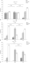
Results: CTA did not differ significantly from DSA (P = .155), and both CTA (P = .001) and DSA (P = .007) performed significantly better than MRA in the population as a whole. Differences in performance among the methods, however, depended upon the segmental location of the fistula along the internal carotid artery (ICA). CTA and MRA were similar in detection of CCFs in patients with a fistula at segment 3. CTA significantly outperformed MRA in patients with a fistula at segment 4, who accounted for approximately half of the population.
Conclusions: CTA source imaging has proved itself as useful as DSA for detecting CCFs. Of the 2 noninvasive techniques, CTA performed better than MRA in the population as a whole and in most patients whose fistula was located at segment 4 or 5 of the ICA.
Figures


Similar articles
-
Natural course of partially embolized carotid-cavernous fistulas.J Neuroimaging. 2024 May-Jun;34(3):376-385. doi: 10.1111/jon.13192. Epub 2024 Feb 11. J Neuroimaging. 2024. PMID: 38343141
-
Comparison of multidetector CT angiography and MR imaging of cervical artery dissection.AJNR Am J Neuroradiol. 2008 Oct;29(9):1753-60. doi: 10.3174/ajnr.A1189. Epub 2008 Jul 17. AJNR Am J Neuroradiol. 2008. PMID: 18635617 Free PMC article.
-
Feasibility of Noninvasive Diagnosis and Treatment Planning in a Case Series with Carotid-Cavernous Fistula using High-Resolution Time-Resolved MR-Angiography with Stochastic Trajectories (TWIST) and Extended Parallel Acquisition Technique (ePAT 6) at 3 T.Clin Neuroradiol. 2015 Sep;25(3):241-7. doi: 10.1007/s00062-014-0298-2. Epub 2014 Mar 6. Clin Neuroradiol. 2015. PMID: 24599323
-
[Carotid-cavernous fistulas].Radiologie (Heidelb). 2024 Mar;64(3):182-188. doi: 10.1007/s00117-024-01269-1. Epub 2024 Feb 13. Radiologie (Heidelb). 2024. PMID: 38351202 Review. German.
-
Endovascular techniques for treatment of carotid-cavernous fistula.J Neuroophthalmol. 2009 Mar;29(1):62-71. doi: 10.1097/WNO.0b013e3181989fc0. J Neuroophthalmol. 2009. PMID: 19458580 Review.
Cited by
-
Susceptibility-weighted imaging in carotido-cavernous fistulas. A case control study.Interv Neuroradiol. 2013 Dec;19(4):438-44. doi: 10.1177/159101991301900406. Epub 2013 Dec 18. Interv Neuroradiol. 2013. PMID: 24355147 Free PMC article.
-
Bilateral Low-Flow Type-D Dural Carotid-Cavernous Fistula: Diagnosis and Treatment with 3D Time-of-Flight Magnetic Resonance Angiography.Am J Case Rep. 2024 Mar 20;25:e942833. doi: 10.12659/AJCR.942833. Am J Case Rep. 2024. PMID: 38504435 Free PMC article.
-
The non-invasive search for the carotid-cavernous fistula: the added value of the 4D-CTA.Neuroradiology. 2017 Sep;59(9):835-837. doi: 10.1007/s00234-017-1885-0. Epub 2017 Jul 25. Neuroradiology. 2017. PMID: 28744728 No abstract available.
-
Transvenous embolization of a carotid-cavernous fistula via the inferior ophthalmic vein: illustrative case.J Neurosurg Case Lessons. 2024 Aug 12;8(7):CASE24183. doi: 10.3171/CASE24183. Print 2024 Aug 12. J Neurosurg Case Lessons. 2024. PMID: 39133941 Free PMC article.
-
Endovascular Management of Intracranial Dural AVFs: Principles.AJNR Am J Neuroradiol. 2022 Feb;43(2):160-166. doi: 10.3174/ajnr.A7304. Epub 2021 Oct 21. AJNR Am J Neuroradiol. 2022. PMID: 34674996 Free PMC article. Review.
References
-
- Modic MT, Berlin AJ, Weinstein MA. The use of digital subtraction angiography in the evaluation of carotid cavernous sinus fistulas. Ophthalmology 1982;89:441–444 - PubMed
-
- Killic T, Elmaci I, Bayri Y, et al. Value of transcranial Doppler ultrasonography in the diagnosis and follow-up of carotid-cavernous fistulae. Acta Neurochir (Wien) 2001;143:1257–1264 - PubMed
-
- Molnar LJ, Caldas JG, Garcia RG, Cerri GG. Doppler mapping of direct carotid-cavernous fistulae (DCCF). Ultrasound Med Biol 2001;27:367–371 - PubMed
Publication types
MeSH terms
Substances
LinkOut - more resources
Full Text Sources
Miscellaneous
