Subarachnoid hemorrhage due to isolated spinal artery aneurysm in four patients
- PMID: 16219857
- PMCID: PMC7976143
Subarachnoid hemorrhage due to isolated spinal artery aneurysm in four patients
Abstract
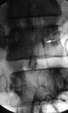
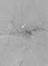
Spinal artery aneurysms are usually found with arteriovenous malformations or other entities that increase hemodynamic stress. Isolated spinal artery aneurysms are rare. Four patients who presented with the acute onset of lower back pain underwent MR imaging, which revealed spinal subarachnoid hemorrhage. In all patients, work-up yielded a diagnosis of isolated spinal aneurysm, and operative treatment was successful. In the appropriate clinical setting, spinal aneurysm should be considered as a possible cause of spinal subarachnoid hemorrhage.
Figures


Comment in
-
Subarachnoid hemorrhage due to isolated spinal arteries: Rare cases with controversy about the treatment strategy.AJNR Am J Neuroradiol. 2006 Apr;27(4):726-7; author reply 727. AJNR Am J Neuroradiol. 2006. PMID: 16611753 Free PMC article. No abstract available.
Similar articles
-
Spontaneous spinal subarachnoid hemorrhage secondary to spinal aneurysms: diagnosis and treatment paradigm.Neurosurgery. 2005 Dec;57(6):1127-31; discussion 1127-31. doi: 10.1227/01.neu.0000186010.34779.10. Neurosurgery. 2005. PMID: 16331160
-
Spontaneous dissecting aneurysms of the basilar artery presenting with a subarachnoid hemorrhage. Report of two cases.J Neurosurg. 1991 Oct;75(4):628-33. doi: 10.3171/jns.1991.75.4.0628. J Neurosurg. 1991. PMID: 1885981 Review.
-
Spinal arterial aneurysm: case report.Neurosurgery. 1993 Jul;33(1):125-9; discussion 129-30. doi: 10.1227/00006123-199307000-00020. Neurosurgery. 1993. PMID: 8355828 Review.
-
Dissection aneurysm of the radiculomedullary branch of the artery of Adamkiewicz with subarachnoid hemorrhage.Neurol Med Chir (Tokyo). 2011;51(9):649-52. doi: 10.2176/nmc.51.649. Neurol Med Chir (Tokyo). 2011. PMID: 21946730
-
Subarachnoid hemorrhage due to anterior spinal artery aneurysm.Neurosurgery. 1986 Feb;18(2):217-9. doi: 10.1227/00006123-198602000-00020. Neurosurgery. 1986. PMID: 3960303
Cited by
-
Spinal subarachnoid hemorrhage due to ruptured solitary aneurysm at thoracolumbar level with fatal outcome.J Neurol. 2007 Jul;254(7):950-2. doi: 10.1007/s00415-006-0288-7. Epub 2007 Apr 20. J Neurol. 2007. PMID: 17446995 No abstract available.
-
Ruptured anterior spinal artery aneurysm from a herniated cervical disc. A case report and review of the literature.Surg Neurol Int. 2016 Jan 28;7:10. doi: 10.4103/2152-7806.175072. eCollection 2016. Surg Neurol Int. 2016. PMID: 26862449 Free PMC article.
-
Subarachnoid hemorrhage due to isolated spinal arteries: Rare cases with controversy about the treatment strategy.AJNR Am J Neuroradiol. 2006 Apr;27(4):726-7; author reply 727. AJNR Am J Neuroradiol. 2006. PMID: 16611753 Free PMC article. No abstract available.
-
Management considerations in ruptured isolated radiculopial artery aneurysms. A report of two cases and literature review.Interv Neuroradiol. 2013 Mar;19(1):60-6. doi: 10.1177/159101991301900109. Epub 2013 Mar 4. Interv Neuroradiol. 2013. PMID: 23472725 Free PMC article. Review.
-
Posterior Spinal Artery Aneurysm Presenting with Leukocytoclastic Vasculitis.J Cerebrovasc Endovasc Neurosurg. 2016 Mar;18(1):42-7. doi: 10.7461/jcen.2016.18.1.42. Epub 2016 Mar 31. J Cerebrovasc Endovasc Neurosurg. 2016. PMID: 27114966 Free PMC article.
References
-
- Vincent FM. Anterior spinal artery aneurysm presenting as a subarachnoid hemorrhage. Stroke 1981;12:230–232 - PubMed
-
- Rengachary SS, Duke DA, Tsai FY, Kragel PJ. Spinal arterial aneurysm: case report. Neurosurgery 1993;33:125–129 - PubMed
-
- Chen CC, Bellon RJ, Ogilvy CS, Putman CM. Aneurysms of the lateral spinal artery: report of two cases. Neurosurgery 2001;48:949–953 - PubMed
-
- Kawamura S, Yoshida T, Nonoyama Y, Yamada M, Suzuki A, Yasui N. Ruptured anterior spinal artery aneurysm: a case report. Surg Neurol 1999;51:608–612 - PubMed
-
- Vishteh AG, Brown AP, Spetzler RF. Aneurysm of the intradural artery of Adamkiewicz treated with muslin wrapping: technical case report. Neurosurgery 1997;40:207–09 - PubMed
Publication types
MeSH terms
LinkOut - more resources
Full Text Sources
