. 2005 Oct;1(2):e11.
doi: 10.1371/journal.ppat.0010011. Epub 2005 Oct 7.

Increased fidelity reduces poliovirus fitness and virulence under selective pressure in mice

Affiliations

Increased fidelity reduces poliovirus fitness and virulence under selective pressure in mice

Julie K Pfeiffer et al. PLoS Pathog. 2005 Oct.

Abstract

RNA viruses have high error rates, and the resulting quasispecies may aid survival of the virus population in the presence of selective pressure. Therefore, it has been theorized that RNA viruses require high error rates for survival, and that a virus with high fidelity would be less able to cope in complex environments. We previously isolated and characterized poliovirus with a mutation in the viral polymerase, 3D-G64S, which confers resistance to mutagenic nucleotide analogs via increased fidelity. The 3D-G64S virus was less pathogenic than wild-type virus in poliovirus-receptor transgenic mice, even though only slight growth defects were observed in tissue culture. To determine whether the high-fidelity phenotype of the 3D-G64S virus could decrease its fitness under a defined selective pressure, we compared growth of the 3D-G64S virus and 3D wild-type virus in the context of a revertible attenuating point mutation, 2C-F28S. Even with a 10-fold input advantage, the 3D-G64S virus was unable to compete with 3D wild-type virus in the context of the revertible attenuating mutation; however, in the context of a non-revertible version of the 2C-F28S attenuating mutation, 3D-G64S virus matched the replication of 3D wild-type virus. Therefore, the 3D-G64S high-fidelity phenotype reduced viral fitness under a defined selective pressure, making it likely that the reduced spread in murine tissue could be caused by the increased fidelity of the viral polymerase.


Conflict of interest statement

Competing interests. The authors have declared that no competing interests exist.

Figures

Figure 1
Figure 1. Pathogenesis of Wild-Type and 3D-G64S Viruses in PVR Mice
PVR mice were given IM injections with 5 × 10⁶ PFU of either the wild-type or 3D-G64S viruses and were monitored for symptoms of disease. Mice were scored as having symptoms when weakness was first observed in the inoculated limb. Asterisks denote time points where statistically significant differences (p < 0.05, determined by two-sample Student's t-test [d 3, 0.0025; d 4, 0.0053; d 5, 0.043]) were observed between 3D-G64S and wild-type viruses. DOI: 10.1371/journal.ppat.0010011.g001
Figure 2
Figure 2. Viral Competition Assay in Poliovirus Receptor-Expressing Mice
(A) Diagram of poliovirus genome showing BstBI restriction enzyme sites within the coding region for 3D polymerase. Digestion of 1,024-bp 32P end-labeled (*) RT-PCR products yields a 558-bp labeled band for wild-type and a 396-bp labeled band for 3D-G64S. 6-wk-old mice were given IM injections with a 1/2 mixture of wild-type to 3D-G64S poliovirus (2 × 10⁷ PFU total virus per mouse). Tissues were harvested when animals first developed paralysis, between days 3 and 5. PCR products were end-labeled with 32P during synthesis by inclusion of a radiolabeled downstream primer and products were digested with BstBI before electrophoresis on a denaturing polyacrylamide gel. Products from muscle samples from the IM infections are shown in (B) while products from brain samples of the IM infections are shown in (C). In (D), 2-wk-old mice were given IC injections with a 1/1 mixture of wild-type to 3D-G64S virus, and brain tissue was harvested when mice became lethargic or paralyzed. For (B), (C), and (D), representative gels are shown. In (E), (F), and (G) all mouse data obtained from the types of experiments shown in (B), (C), and (D), respectively, are quantified, and show the total number of mice whose tissues contained predominately wild-type virus, 3D-G64S virus, or both. The total number of mice per condition are as follows: IM-muscle samples, 31 mice; IM-brain samples, 54 mice; IC-brain samples, 10 mice. DOI: 10.1371/journal.ppat.0010011.g002
Figure 3
Figure 3. Growth of Wild-Type and 3D-G64S Poliovirus in HeLa Cells
(A) Single-cycle growth curve. 2 × 10⁶ HeLa cells were infected at an MOI of 10 PFU/cell with either wild-type or 3D-G64S viruses at 37 °C. Cell-associated virus was harvested at indicated times and titered by plaque assay. (B) Plaque assays. Wild-type or 3D-G64S poliovirus was plated on HeLa cell monolayers and incubated under an agar overlay for 48 h at indicated temperatures before staining the cells with crystal violet. (C) Competition assay titration with plasmid DNA. Plasmid DNAs that encode wild-type and 3D-G64S poliovirus were diluted and mixed at the ratios indicated: for example, 1/100 denotes a ratio of 0.1 ng of wild-type viral cDNA to 10 ng of 3D-G64S cDNA. End-labeled PCR products were digested with BstBI and were electrophoresed on denaturing polyacrylamide gels. (D) Competition assay in HeLa cells infected with wild-type and 3D-G64S virus. HeLa cells (2 × 10⁶) were infected at an MOI of 0.1 or 10 PFU/cell, as indicated, with wild-type virus, 3D-G64S virus, or both, and cells were harvested 5 h after infection. RNA was extracted, subjected to RT-PCR as in (C), and digested with BstBI. Control bands “uncut” and “mix” were amplified from cDNAs. Products from MOI 0.1 single infections with wild-type, 3D-G64S- and mock-infected cells are shown, as are mixed infections with a 1/1 ratio of wild-type and 3D-G64S virus (MOI of 0.1 PFU/cell or 10 PFU/cell for each virus) and a 1/2 ratio (MOI of 5 and 10 PFU/cell, respectively). Products were quantified by Phosphorimager and the percentage 3D-G64S is shown. DOI: 10.1371/journal.ppat.0010011.g003
Figure 4
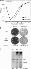
Figure 4. Growth of Wild-Type and 3D-G64S Poliovirus in Primary PVR-MEF
(A) Single-cycle growth curve. 2 × 10⁵ PVR-MEF were infected at an MOI of 10 PFU/cell with either wild-type or 3D-G64S viruses at 37 °C. Cell-associated virus was harvested at indicated times and titered by plaque assay. (B) Plaque assays. Wild-type or 3D-G64S poliovirus was plated on HeLa or PVR-MEF monolayers and incubated under an agar overlay for 72 h at 37 °C before staining the cells with crystal violet. The amount of virus plated, according to HeLa titration, is shown underneath as “input.” (C) Serial passage competition assay in PVR-MEF. 3D-G64S and wild-type virus were mixed in a 1:1 ratio and used to infect PVR-MEF at an MOI of 10 PFU/cell at 37 °C. At 6 h post-infection, cell-associated RNA and virus were harvested, and the virus was used to infect fresh cells at high MOI (5–30 PFU/cell). This cycle was repeated a total of three times; BstBI digestion assay products are shown for passages 1, 2, and 3 (P1, P2, P3), and the Phosphorimager-quantified percentage of the G64S product is shown. DOI: 10.1371/journal.ppat.0010011.g004
Figure 5
Figure 5. Serial Passage Competition Assay in HeLa Cells for Mixtures of Wild-Type and 3D-G64S Viruses Containing the 2C-F28S Temperature-Sensitive Attenuating Mutation
One-to-one mixtures of 3D-G64S to WT virus containing the indicated 2C-F28S mutations were used to infect HeLa cells at an MOI of 0.1 PFU/cell at 32 °C. At 12 h post-infection, cell-associated RNA and virus were harvested, and the virus was titered to determine input for the next passage. This cycle was repeated three to five times, and representative gels of digestion products, with maps indicating the mutations at the 2C locus in the mixtures of viruses that were wild-type and 3D-G64S at the 3D locus, are shown: (A) wild-type, (B) 2C-F28S(1m), (C) 2C-F28S(3m). X denote the numbers of nucleotide substitutions at the 2C-F28S site, while O denotes the presence of the 3D-G64S mutation. DOI: 10.1371/journal.ppat.0010011.g005
Figure 6
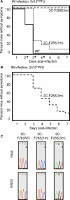
Figure 6. Pathogenesis of Mixtures of Wild-Type and 3D-G64S Viruses Containing the 2C-F28S Temperature-Sensitive Attenuating Mutation
PVR mice were injected intramuscularly with 5 × 10⁶ (A) or 2 × 10⁸ (B) PFU of total virus. The viral inocula were 1/10 mixtures of the wild-type/3D-G64S derivatives to allow 3D-G64S viruses a chance to compete. For example, 2C-F28S(1m) was a mixture of 5 × 10⁵ PFU of 2C-F28S(1m) and 4.5 × 10⁶ PFU of 2C-F28S(1m)/3D-G64S, giving a total of 5 × 10⁶ PFU. The numbers of mice in each group, as designated by the identity of the 2C allele, were: wild-type, 26 mice; 2C-F28S(1m), 32 mice; 2C-F28S(3m), 4 and 27 mice in (A) and (B), respectively. Asterisks denote time points where statistically significant differences (p < 0.05, determined by two-sample Student's t-test, [d 2, 0.00125; d 3, 0.00125; d 4, 0.0112]) were observed between 2C-F28S(1m) and wild-type viruses. (C) Sequence analysis of RT-PCR products from viral RNA pools (“input”) used to inoculate the mice shown in (A) and from muscle tissue after development of disease (“output”). For output samples, samples from at least three mice were sequenced, and a representative example for each is shown. DOI: 10.1371/journal.ppat.0010011.g006
Figure 7
Figure 7. Viral Competition Assay in the Context of the 2C-F28S Temperature-Sensitive Attenuating Mutations
Muscle tissue from the experiment shown in Figure 6 was processed and the BstBI digestion assay was performed. Representative gels of digestion products, with maps indicating the mutations at the 2C locus are shown: (A) wild-type, (B) 2C-F28S(1m), (C) 2C-F28S(3m). (D) Quantitation of the gels in A–C. The percent 3D-G64S is shown, and represents an average from 5–10 mice for each condition. DOI: 10.1371/journal.ppat.0010011.g007

References

    1. Domingo E, Holland JJ. A virus mutations and fitness for survival. Annu Rev Microbiol. 1997;51:151–178. - PubMed
    1. Drake JW, Charlesworth B, Charlesworth D, Crow JF. Rates of spontaneous mutation. Genetics. 1998;148:1667–1686. - PMC - PubMed
    1. Crotty S, Maag D, Arnold JJ, Zhong W, Lau JY, et al. The broad-spectrum antiviral ribonucleoside ribavirin is an RNA virus mutagen. Nat Med. 2000;6:1375–1379. - PubMed
    1. Domingo E, Menendez-Arias L, Holland JJ. RNA virus fitness. Rev Med Virol. 1997;7:87–96. - PubMed
    1. Crotty S, Cameron CE, Andino R. RNA virus error catastrophe: Direct molecular test by using ribavirin. Proc Natl Acad Sci U S A. 2001;98:6895–6900. - PMC - PubMed

Publication types

Associated data