Kidney tubular epithelium is restored without replacement with bone marrow-derived cells during repair after ischemic injury
- PMID: 16221175
- PMCID: PMC2915580
- DOI: 10.1111/j.1523-1755.2005.00629.x
Kidney tubular epithelium is restored without replacement with bone marrow-derived cells during repair after ischemic injury
Abstract
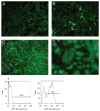
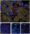
The kidney has the ability to restore the structural and functional integrity of the proximal tubule, which undergoes extensive epithelial cell death after prolonged exposure to ischemia. In order to study the role that adult bone marrow-derived stem cells might play in kidney remodeling after injury, we employed a murine model of ischemia/reperfusion (I/R) injury in which the degree of injury, dysfunction, repair, tubular cell proliferation and functional recovery have been characterized [Park KM, et al, J Biol Chem 276:11870-11876, 2001]. We generated chimeric mice using marrow from mice expressing the bacterial LacZ gene, or the enhanced green fluorescence protein (eGFP) gene, or from male mice transplanted into female mice. The establishment of chimerism was confirmed at 6 weeks following transplantation in each case. I/R injury was induced in chimeric mice by occluding the renal arteries and veins with microaneurysm clamps for 30 minutes. After functional recovery in the eGFP chimeras, although there were many interstitial cells, no tubular cells were derived from bone marrow cells. In the bacterial beta-galactosidase (beta-gal) chimeric mice we found evidence of mammalian (endogenous) beta-gal by 5-bomo-4-chloro-3-indolyl-beta-D-galactopyranoside (X-gal) staining, but not bacterial beta-gal in tubule cells. Detection of the Y chromosome by fluorescence in situ hybridization (FISH) in the postischemic kidneys of gender-mismatched chimeras revealed Y chromosome positivity only in the nuclei of interstitial cells, when scrutinized by deconvolution microscopy. In our model of I/R injury there was a large amount of proliferation of surviving, injured tubular cells indicating that the injured tubule is repopulated by daughter cells of surviving tubular cells. Analysis of the phenotype of interstitial and vascular cells following I/R injury revealed small numbers of peritubular endothelial cells to be derived from bone marrow cells that may serve in the repair process.
Figures


Comment in
-
Could tubular interstitium be a source of adult epithelial stem cells?Kidney Int. 2006 Dec;70(11):2040; author reply 2040-1. doi: 10.1038/sj.ki.5001842. Kidney Int. 2006. PMID: 17130826 No abstract available.
References
-
- Orlic D, Kajstura J, Chimenti S, et al. Transplanted adult bone marrow cells repair myocardial infarcts in mice. Ann NY Acad Sci. 2001;938:221–229. [discussion 229–230] - PubMed
-
- Krause DS, Theise ND, Collector MI, et al. Multi-organ, multi-lineage engraftment by a single bone marrow-derived stem cell. Cell. 2001;105:369–377. - PubMed
-
- Lagasse E, Connors H, Al-Dhalimy M, et al. Purified hematopoietic stem cells can differentiate into hepatocytes in vivo. Nat Med. 2000;6:1229–1234. - PubMed
-
- Ferrari G, Cusella-De Angelis G, Coletta M, et al. Muscle regeneration by bone marrow-derived myogenic progenitors. Science. 1998;279:1528–1530. - PubMed
-
- Poulsom R, Forbes SJ, Hodivala-Dilke K, et al. Bone marrow contributes to renal parenchymal turnover and regeneration. J Pathol. 2001;195:229–235. - PubMed
Publication types
MeSH terms
Grants and funding
LinkOut - more resources
Full Text Sources
Other Literature Sources

