. 2005 Oct 25;102(43):15569-74.
doi: 10.1073/pnas.0507257102. Epub 2005 Oct 12.

Different cell surface oligomeric states of B7-1 and B7-2: implications for signaling

Affiliations

Different cell surface oligomeric states of B7-1 and B7-2: implications for signaling

Sumeena Bhatia et al. Proc Natl Acad Sci U S A. .

Abstract

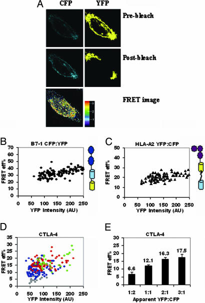
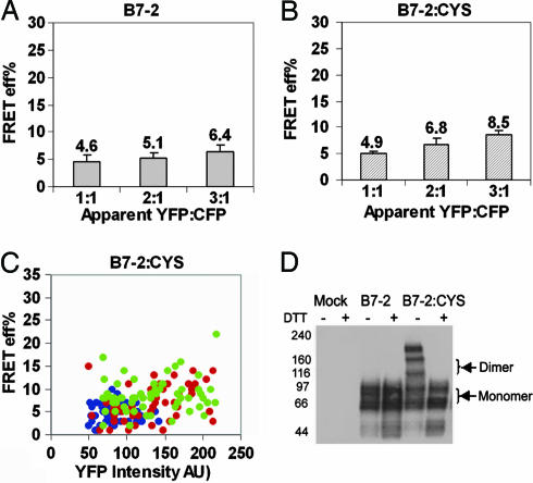
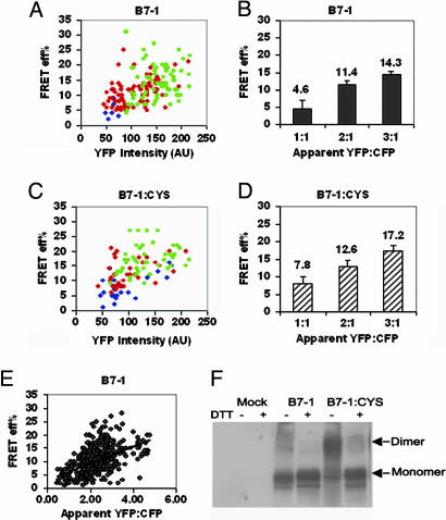
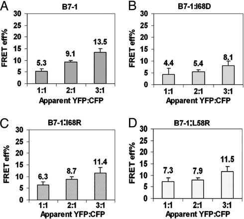
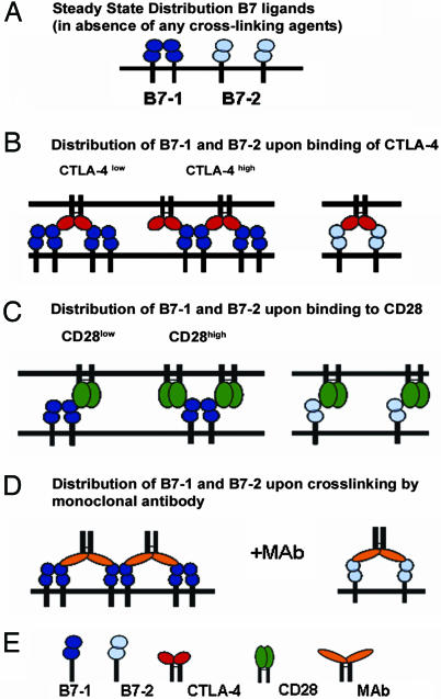
The costimulatory ligands B7-1 and B7-2 are expressed on the surface of antigen-presenting cells and interact with the costimulatory receptors CD28 and cytotoxic T lymphocyte-associated antigen 4 (CTLA-4) expressed on T cells. Although B7-1 and B7-2 are homologous ligands having common receptors, they exhibit distinct biochemical features and roles in immune regulation. Several biochemical and structural studies have indicated differences in the oligomeric state of B7-1 and B7-2. However, the organization of B7 ligands on the cell surface has not been examined. By using photobleaching-based FRET (pbFRET), we demonstrate that B7-1 and B7-2 adopt different oligomeric states on the cell surface. Our study shows that B7-2 exists as a monomer on the cell surface whereas B7-1 exists predominantly as dimers on the cell surface. A series of mutations in B7-1 result in the expression of a predominantly monomeric species on the cell surface and validate the dimer interface proposed by prior crystallographic analysis. The difference in the oligomeric states of B7-1 and B7-2 provides insight into the geometric organization of the costimulatory receptor-ligand complexes in the immunological synapse and suggests constraints on signal transduction mechanisms involved in T cell activation.


Figures

Fig. 1.
Fig. 1.
Validation of pbFRET for covalently linked CFP and YFP tandem proteins and characterization of CTLA-4. (A) Pre- and postbleaching of cells expressing CFP and YFP (Top and Middle row; note the enhancement in the intensity of CFP postbleaching). (Bottom) The pseudocolored FRET image with a color gradient from blue to red with values 1 (minimum) to 3 (maximum) for FRET. FRET efficiency, E, for tandem (B7-1)-CFP-YFP (B) and tandem (HLA-A2)-YFP-CFP (C) is independent of YFP intensity, over a wide range of YFP intensities, expressed as arbitrary units (AU). (D) E for CTLA-4 is independent of YFP intensity at each acceptor (YFP) to donor (CFP) ratio: gray (1:2), blue (1:1), red (2:1). and green (3:1) YFP-to-CFP ratio. (E) Average E for CTLA-4 increases with an increase in YFP-to-CFP ratio. The average E was calculated from the E corresponding to the range of 50–220 AUs of YFP to exclude the very low and very high intensity points that either give rise to noise or are outside the experimentally observed range. The error bars represent the 0.01% confidence limits. The P values between the adjacent ratios of YFP to CFP (i.e., 1:2 and 1:1, 1:1 and 2:1) were <0.01. The data are pooled from three independent experiments.
Fig. 2.
Fig. 2.
B7-2 as a monomer on the cell surface. (A) Average E (calculated as in Fig. 1E) for B7-2 does not change with an increase in YFP-to-CFP ratio. (B) Average E for B7-2:CYS does not show a consistently significant increase with increase in YFP-to-CFP ratio. (C) E for B7-2:CYS modestly increases with an increase in YFP intensity at different acceptor-to-donor ratios; 1:1 (blue), 2:1 (red), and 3:1 (green) YFP-to-CFP ratios. The data are pooled from two independent experiments. The P value between the adjacent ratios of YFP to CFP in A was >0.05. For B, the P value was <0.01 between 1:1 and 2:1 and >0.01 between 2:1 and 3:1 YFP-to-CFP ratio. The error bars in A and B represent the 0.01% confidence limits. (D) Whole cell lysates of CHO cells transiently expressing B7-2-YFP or B7-2:CYS-YFP were immunoblotted with anti-B7-2. Western blot analysis shows that B7-2:CYS exists as a mixture of monomers and dimers. The “artifactual” dimers of B7-2:CYS arise from cells expressing very high levels of B7-2:CYS.
Fig. 3.
Fig. 3.
B7-1 is predominantly a dimer on the cell surface. (A) E for B7-1 increases with an increase in YFP intensity at each YFP-to-CFP ratio. (B) Average E (calculated as in Fig. 1E) increases with an increase in YFP-to-CFP ratio. (C) E for B7-1:CYS increases with an increase in YFP intensity at each YFP-to-CFP ratio. (D) Average E for B7-1:CYS increases with an increase in YFP-to-CFP ratio. The symbols in A and C are 1:1 (blue), 2:1 (red), and 3:1 (green) YFP-to-CFP ratios. The error bars in B and D represent the 0.01% confidence limits. The data are representative of two independent experiments. The P value between the adjacent ratios of YFP to CFP in B and D was <0.01. (E) E for B7-1 shows a linear dependence on YFP-to-CFP ratio. (F) Western blot of whole cell lysates of cells transiently expressing B7-1 or B7-1:CYS shows the presence of dimers of B7-1:CYS.
Fig. 4.
Fig. 4.
Mutations at L58 and I68 residues interfere with the dimeric state of B7-1. (A) E for WT B7-1 increases with an increase in YFP-to-CFP ratio whereas E for I68D (B), I68R (C), and L58R (D) does not show any systematic dependence on YFP-to-CFP ratio. The error bars represent the 0.01% confidence limits. The data are pooled from three independent experiments. The P value between 1:1 and 2:1 YFP-to-CFP ratio in B, C, and D was ≈0.5 whereas in A it was <0.01.
Fig. 5.
Fig. 5.
Models for the interaction of CTLA-4 and CD28 with B7-1 and B7-2. (A) Steady-state distribution of B7-1 (dimer) and B7-2 (monomer). (B) Assemblies of B7-1 and B7-2 upon binding to CTLA-4. At low concentration of CTLA-4 (relative to B7-1), a bivalent homodimeric CTLA-4 molecule can bind two B7-1 molecules, but, at high concentration of CTLA-4, it may form an extended array with B7-1. However, with monomeric B7-2, CTLA-4 can engage only two B7-2 monomers. (C) Assemblies of B7-1 and B7-2 upon binding to CD28. At low concentration of monovalent CD28, a dimeric B7-1 could engage a single CD28 molecule; however, at high concentration of CD28, two molecules of CD28 may be bridged by one molecule of B7-1 without the possibility of forming higher order assemblies. With monomeric B7-2, monovalent CD28 may form single solitary complexes. (D) Assemblies of B7-1 and B7-2 upon cross-linking by bivalent monoclonal antibodies. Anti-B7-1 antibodies may interact with dimeric B7-1 like CTLA-4 and form an ordered network whereas anti-B7-2 antibodies may simply induce bridging of two B7-2 monomers. (E) Key for the symbols.

References

    1. Peach, R. J., Bajorath, J., Brady, W., Leytze, G., Greene, J., Naemura, J. & Linsley, P. S. (1994) J. Exp. Med. 180, 2049–2058. - PMC - PubMed
    1. Greene, J. L., Leytze, G. M., Emswiler, J., Peach, R., Bajorath, J., Cosand, W. & Linsley, P. S. (1996) J. Biol. Chem. 271, 26762–26771. - PubMed
    1. van der Merwe, P. A. & Davis, S. J. (2003) Annu. Rev. Immunol. 21, 659–684. - PubMed
    1. Thompson, C. B., Lindsten, T., Ledbetter, J. A., Kunkel, S. L., Young, H. A., Emerson, S. G., Leiden, J. M. & June, C. H. (1989) Proc. Natl. Acad. Sci. USA 86, 1333–1337. - PMC - PubMed
    1. Linsley, P. S., Brady, W., Grosmaire, L., Aruffo, A., Damle, N. K. & Ledbetter, J. A. (1991) J. Exp. Med. 173, 721–730. - PMC - PubMed

Publication types

LinkOut - more resources