. 2005 Oct 25;102(43):15418-22.
doi: 10.1073/pnas.0506750102. Epub 2005 Oct 12.

Coevolution of tRNA intron motifs and tRNA endonuclease architecture in Archaea

Affiliations

Coevolution of tRNA intron motifs and tRNA endonuclease architecture in Archaea

Giuseppe D Tocchini-Valentini et al. Proc Natl Acad Sci U S A. .

Abstract

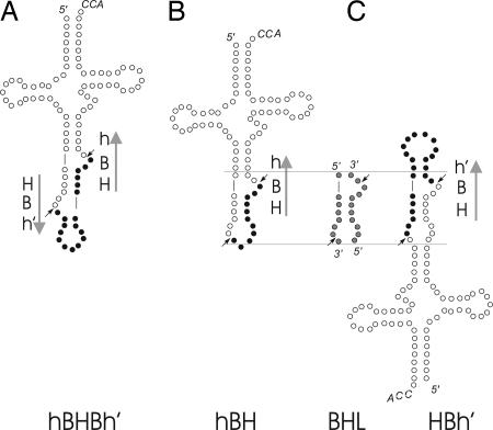
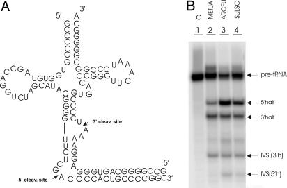
Members of the three kingdoms of life contain tRNA genes with introns. The introns in pre-tRNAs of Bacteria are self-splicing, whereas introns in archaeal and eukaryal pre-tRNAs are removed by splicing endonucleases. We have studied the structures of the endonucleases of Archaea and the architecture of the sites recognized in their pre-tRNA substrates. Three endonuclease structures are known in the Archaea: a homotetramer in some Euryarchaea, a homodimer in other Euryarchaea, and a heterotetramer in the Crenarchaeota. The homotetramer cleaves only the canonical bulge-helix-bulge structure in its substrates. Variants of the substrate structure, termed bulge-helix-loops, appear in the pre-tRNAs of the Crenarcheota and Nanoarcheota. These variant structures can be cleaved only by the homodimer or heterotetramer forms of the endonucleases. Thus, the structures of the endonucleases and their substrates appear to have evolved together.


Figures

Fig. 1.
Fig. 1.
tRNA intron motifs and enzyme architectures. Vertical bars indicate the species sharing the same endonuclease architecture (11). α4 refers to the homotetramer, α2 refers to the homodimer, and α2β2 refers to the heterotetramer (11). The numbers indicate the number of hBHBh (BHB) and HBh or hBH (BHL) motifs present in the genome, according to ref. .
Fig. 2.
Fig. 2.
Archaeal endonucleases common fold. (A) Cartoon representation of the monomeric subunit of METJA (16). All of the side chains represented are conserved among all of the subunits of archaeal endonucleases. The residues constituting the two signatures, as specified in the text, are colored blue and purple; the other conserved residues are yellow. (B) Helical wheel representation of helix α2. Hydrophobic residues are shaded in gray; only the conserved amino acids are numbered. (C) Helical wheel representation of helix α4. Representation as above.
Fig. 3.
Fig. 3.
Canonical and noncanonical motifs. (A) hBHBh′, BHB splicing motif flanked by two helices (h, h′) presenting at least two Watson–Crick base pairs. Arrows indicate the 5′ 32 → 3′ sense. (B and C) hBH and HBh′ relaxed BHB motifs. The two superimposed motifs are shaded in gray to show the similarity to a BHL motif.
Fig. 4.
Fig. 4.
In vitro cleavage of a BHB-containing substrate. (A) The pre-tRNA Archeuka was constructed by using two regions derived from yeast pre-tRNAPhe (nucleotides 1–31 and 38–76) joined by a 25-nt insert that corresponds to the BHB motif of archaeal tRNA Trp. (B) The pre-tRNA Archeuka was incubated with four different enzymes. The conditions of the reactions are reported in Materials and Methods. The cleavage products were analyzed by electrophoresis on 10% polyacrylamide gel containing 29:1 monomer to bis and 8 M Urea, followed by autoradiography. The identification of the reaction products is indicated. Lane 1 is control (C, no enzyme added); lanes 2–5 show the products after incubation with the endonucleases from METJA, SULSO, ARCFU, and XENLA, respectively. The 2/3 molecules are produced by single cleavage. The slowly migrating class corresponds to intron–3′ exon molecules and the fast migrating class to 5′ exon–intron molecules. The heterogeneity of the 3′ halves results from the run-off transcription by T7 RNA polymerase, used to prepare the substrate.
Fig. 5.
Fig. 5.
In vitro cleavage of a BHL-containing substrate. (A) The pre-tRNATyr from Caenorhabditis elegans. The synthetic substrate presents a residue change at the 5′ terminus (C to G) required for T7 transcription and a corresponding change (G to C) in the complementary strand. (B) The pre-tRNATyr was incubated with four different enzymes. The conditions of the reactions and product analysis were the same as described in the legend to Fig. 3.
Fig. 6.
Fig. 6.
In vitro cleavage of a substrate presenting a trans BHL. (A) Proposed structure of the N. equitans pre-tRNAGlu formed by annealing the products of two half-genes. The left side of the structure contains the 5′ half-gene, including the 5′ cleavage site in the loop formed by CGAC. The right side of the structure contains the 3′ cleavage site comprising the AAU bulge. The long GC-rich stem at the bottom of the structure is not covalently closed but that does not matter, because it is removed by the splicing endonuclease. (B) Cleavage of the pre-tRNAGlu by purified archaeal endonucleases. The substrate was incubated with three different recombinant proteins. The cleavage products were analyzed as described in the legend to Fig. 3. The identification of the reaction products is indicated. Lane 1 is control (C, no enzyme added); lanes 2–4 show the products after incubation with the endonucleases from METJA, ARCFU, and SULSO, respectively. Because the substrate molecule is open at the bottom, the enzymes produce, in addition to 5′ and 3′ halves, two intron fragments. IVS (3′h) is the fragment resulting from cleavage at the 3′ cleavage site, and IVS (5′h) is the fragment resulting from cleavage at the 5′ cleavage site.

References

    1. Kjems, J., Jensen, J., Olesen, T. & Garrett, R. A. (1989) Can J. Microbiol. 35, 210-214. - PubMed
    1. Kjems, J. & Garrett, R. A. (1991) Proc. Natl. Acad. Sci. USA 88, 439-443. - PMC - PubMed
    1. Kjems, J. & Garrett, R. A. (1988) Cell 54, 693-703. - PubMed
    1. Tang, T. H., Rozhdestvensky, T. S., d'Orval, B. C., Bortolin, M. L., Huber, H., Charpentier, B., Branlant, C., Bachellerie, J. P., Brosius, J. & Huttenhofer, A. (2002) Nucleic Acids Res. 30, 921-930. - PMC - PubMed
    1. Fabbri, S., Fruscoloni, P., Bufardeci, E., Di Nicola Negri, E., Baldi, M. I., Attardi, D. G., Mattoccia, E. & Tocchini-Valentini, G. P. (1998) Science 280, 284-286. - PubMed

Publication types

LinkOut - more resources