Comparative Study
. 2005 Oct 12;25(41):9339-46.
doi: 10.1523/JNEUROSCI.2282-05.2005.

Formation of a motor memory by action observation

Affiliations
Comparative Study

Formation of a motor memory by action observation

Katja Stefan et al. J Neurosci. .

Abstract

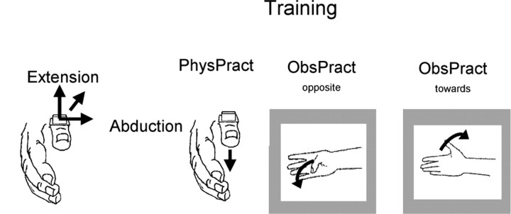
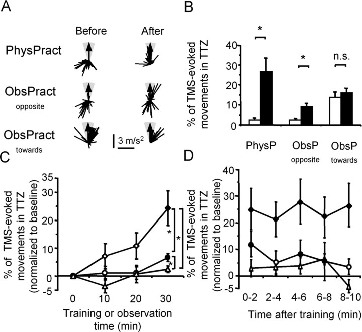
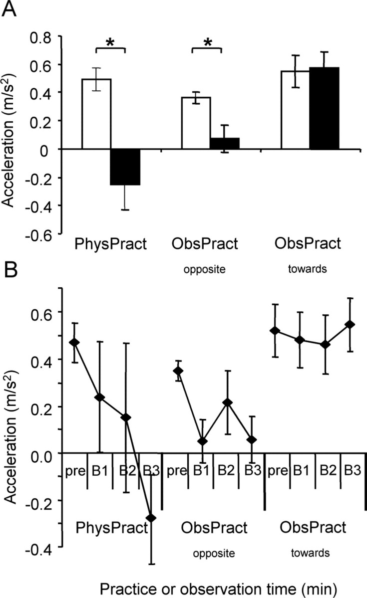
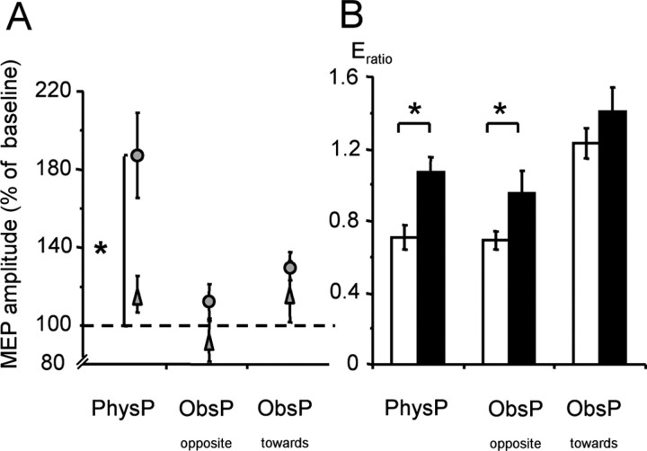
Mirror neurons discharge with both action observation and action execution. It has been proposed that the mirror neuron system is instrumental in motor learning. The human primary motor cortex (M1) displays mirror activity in response to movement observation, is capable of forming motor memories, and is involved in motor learning. However, it is not known whether movement observation can lead directly to the formation of motor memories in the M1, which is considered a likely physiological step in motor learning. Here, we used transcranial magnetic stimulation (TMS) to show that observation of another individual performing simple repetitive thumb movements gives rise to a kinematically specific memory trace of the observed motions in M1. An extended period of observation of thumb movements that were oriented oppositely to the previously determined habitual directional bias increased the probability of TMS-evoked thumb movements to fall within the observed direction. Furthermore, the acceleration of TMS-evoked thumb movements along the principal movement axis and the balance of excitability of muscle representations active in the observed movements were altered in favor of the observed movement direction. These findings support a role for the mirror neuron system in memory formation and possibly human motor learning.


Figures

Figure 1.
Figure 1.
Experimental design. At the beginning of each session, the direction of TMS-evoked movements (“baseline before training”) was determined in each individual by assessing the first-peak accelerations along the extension/flexion and abduction/adduction axes (Classen et al., 1998). Subsequently, subjects participated in three different training sessions: physical training, which consisted of performance of voluntary thumb movements in a direction opposite the baseline direction and observation of thumb movements directed opposite the baseline direction and toward the baseline direction.
Figure 2.
Figure 2.
Effect of different training interventions on TMS-evoked movement direction. A, Example of one subject. First-peak acceleration vectors of TMS-evoked thumb movements before (left) and after (right) each of the three interventions (PhysPract, ObsPractopposite, and ObsPracttoward). For better comparability, all examples are aligned to the training direction indicated by the arrow. The TTZ is shown in gray. PhysPract and ObsPractopposite, but not ObsPracttoward, led to substantial changes in the direction of TMS-evoked movements. B, Group data (n = 10) showing the PTTZ before and after PhysPract, ObsPractopposite, and ObsPracttoward. Before the training interventions, the percentage of TMS-evoked movements in TTZ was similar in PhysPract and ObsPractopposite. PhysPract and ObsPractopposite, but not ObsPracttoward, led to a significant increase in the percentage of TMS-evoked movements falling into TTZ. Data show means ± SEM. *p < 0.005. n.s., Not significant. C, Time course of changes of PTTZ as a function of training intervention. To compare between conditions, ΔPTTZ was computed as the baseline-normalized, intervention-dependent change in percentage of TMS-evoked thumb movements falling into the TTZ (see Materials and Methods). At the end of 30 min, the percentage change of TMS-evoked movements in TTZ was larger in PhysPract than in ObsPractopposite or ObsPracttoward and with ObsPractopposite than with ObsPracttoward. Rhombi, PhysPract; circles, ObsPractopposite; triangles, ObsPracttoward. Filled symbols, Time points significantly different from baseline; two-tailed t tests; false discovery rate correction. *p < 0.05. D, Duration of changes of PTTZ as a function of training intervention. Symbols as in C. Error bars represent SEM.
Figure 3.
Figure 3.
Effect of different training interventions on compound acceleration vector. A, Group data (n = 10) showing the mean compound acceleration vector for the extension/flexion direction before and after training. Before training, the compound acceleration vector of each subject was aligned such that all vectors pointed into the extension direction. After PhysPract, the mean compound acceleration vector came to point into the flexion direction, corresponding to the practiced direction. ObsPractopposite decreased the length of the mean compound acceleration vector, whereas ObsPracttoward did not change the mean compound acceleration vector. *p < 0.01. Open bars, Before training; filled bars, after training. B, Time course of changes of the mean compound acceleration vector as a function of training intervention. At the end of 30 min, the mean compound acceleration vector was smaller in PhysPract than in ObsPractopposite or ObsPracttoward and with ObsPractopposite than with ObsPracttoward. Error bars represent SEM. pre, Before training; B1, B2, and B3, results obtained after the first, second, and third training block, respectively.
Figure 4.
Figure 4.
Effect of different training interventions on corticomuscular excitability. A, Changes in MEP amplitudes (relative to baseline) recorded from training agonist muscle (MEPagonist; circles) and antagonist muscle (MEPantagonist; triangles). PhysPract led to a significant increase in MEPagonist compared with baseline. B, The ratio between the MEPagonist and MEPantagonist increased both after PhysPract and ObsPractopposite but not after ObsPracttoward. Open bars, Before training; filled bars, after training. *p < 0.05. Error bars represent SEM.

References

    1. Astafiev SV, Stanley CM, Shulman GL, Corbetta M (2004) Extrastriate body area in human occipital cortex responds to the performance of motor actions. Nat Neurosci 7: 542-548. - PubMed
    1. Avikainen S, Forss N, Hari R (2002) Modulated activation of the human SI and SII cortices during observation of hand actions. NeuroImage 15: 640-646. - PubMed
    1. Baldissera F, Cavallari P, Craighero L, Fadiga L (2001) Modulation of spinal excitability during observation of hand actions in humans. Eur J Neurosci 13: 190-194. - PubMed
    1. Black CB, Wright DL (2000) Can observational practice facilitate error recognition and movement production? Res Q Exerc Sport 71: 331-339. - PubMed
    1. Brass M, Bekkering H, Prinz W (2001) Movement observation affects movement execution in a simple response task. Acta Psychol (Amst) 106: 3-22. - PubMed

Publication types