Comparative Study
. 2005 Oct 12;25(41):9470-8.
doi: 10.1523/JNEUROSCI.2826-05.2005.

Q/R site editing controls kainate receptor inhibition by membrane fatty acids

Affiliations
Comparative Study

Q/R site editing controls kainate receptor inhibition by membrane fatty acids

Timothy J Wilding et al. J Neurosci. .

Abstract

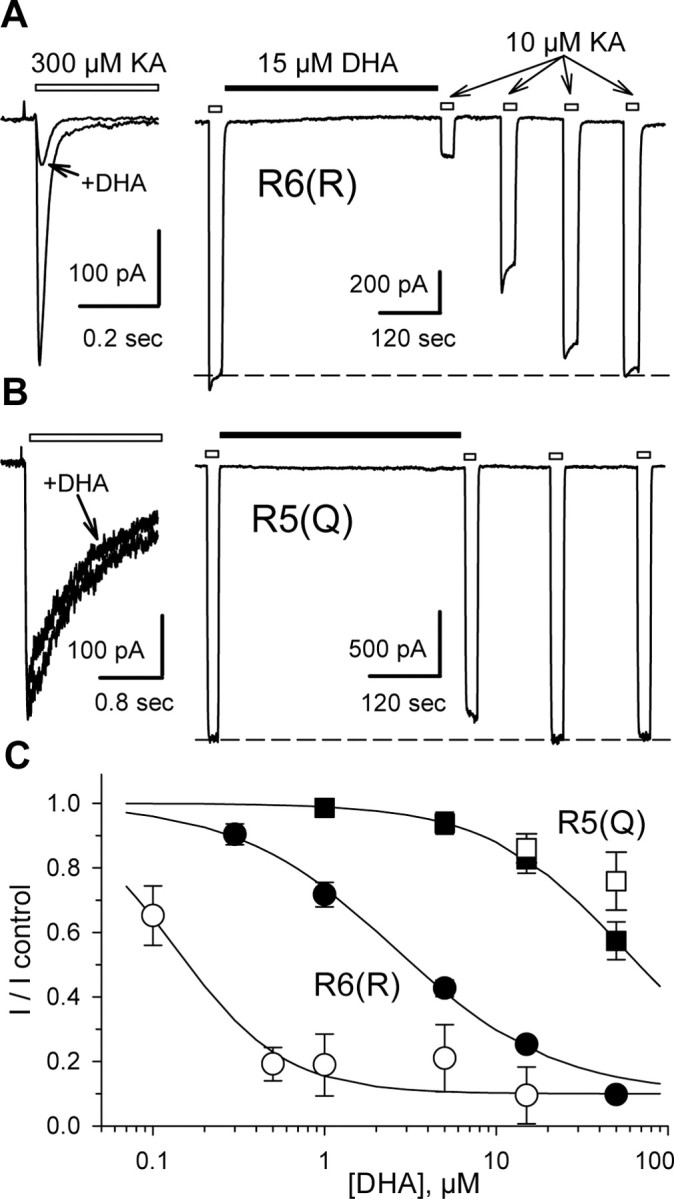
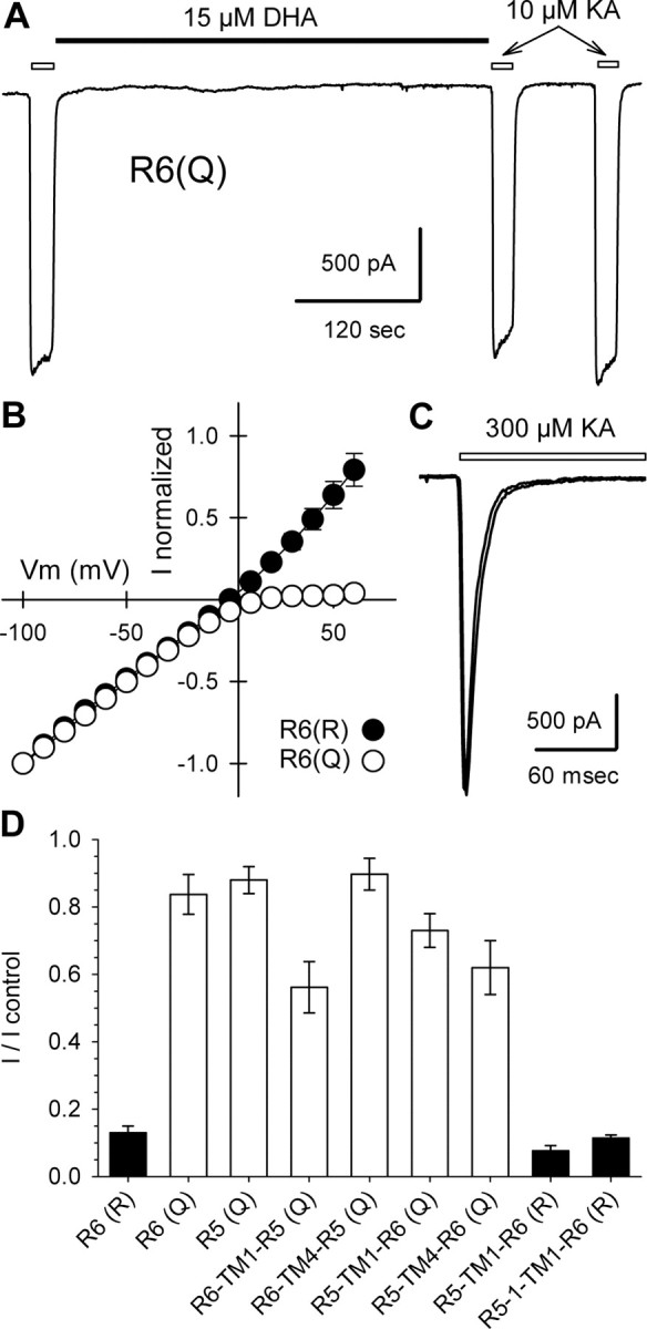
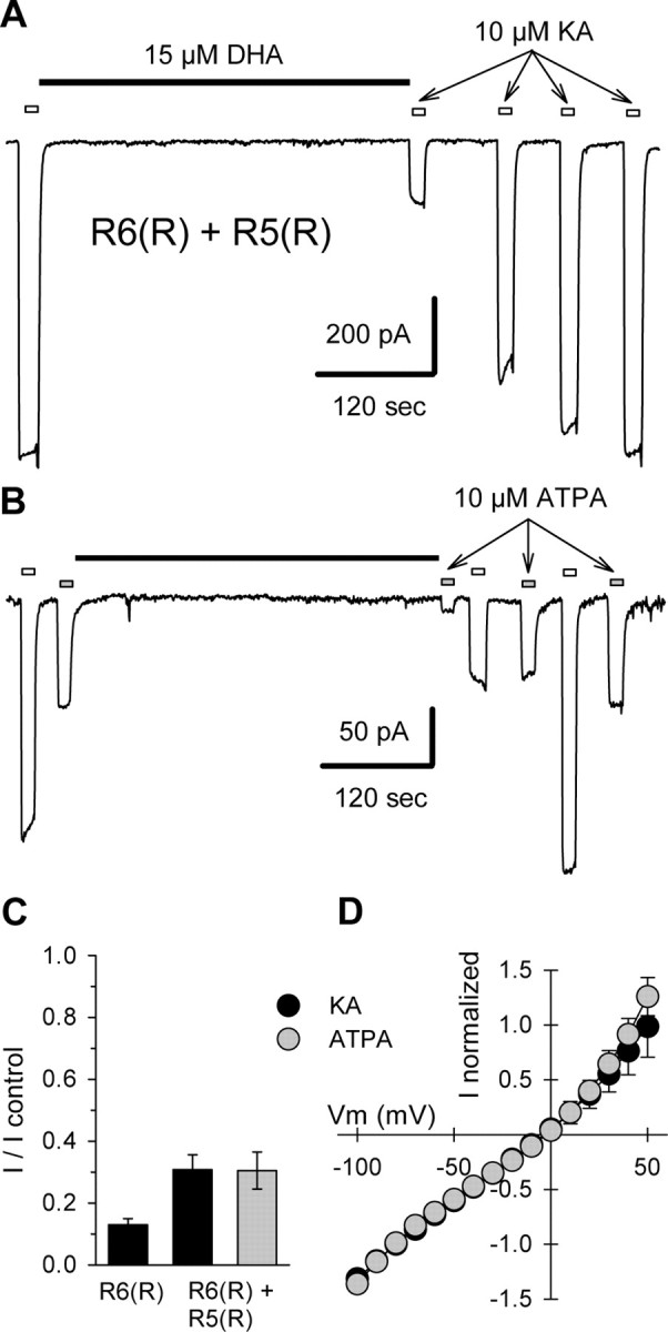
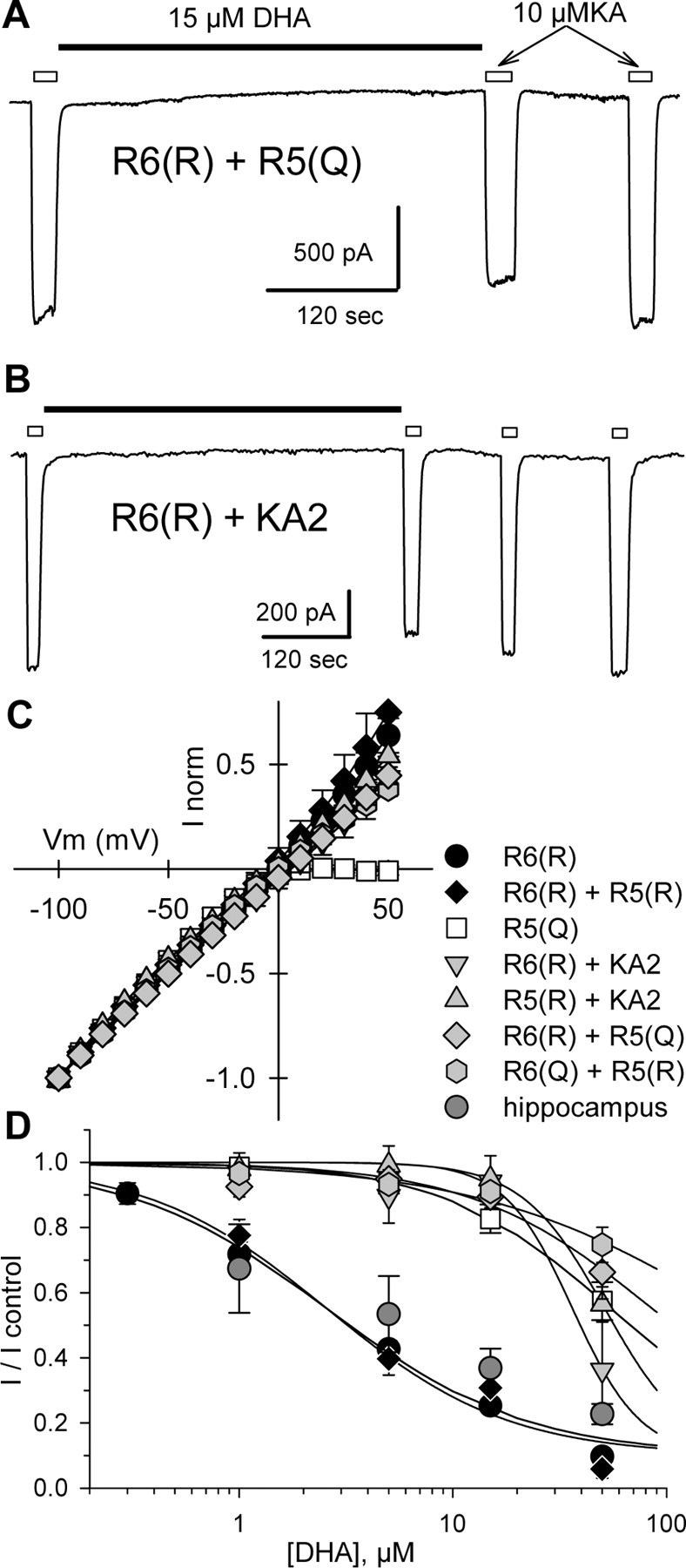
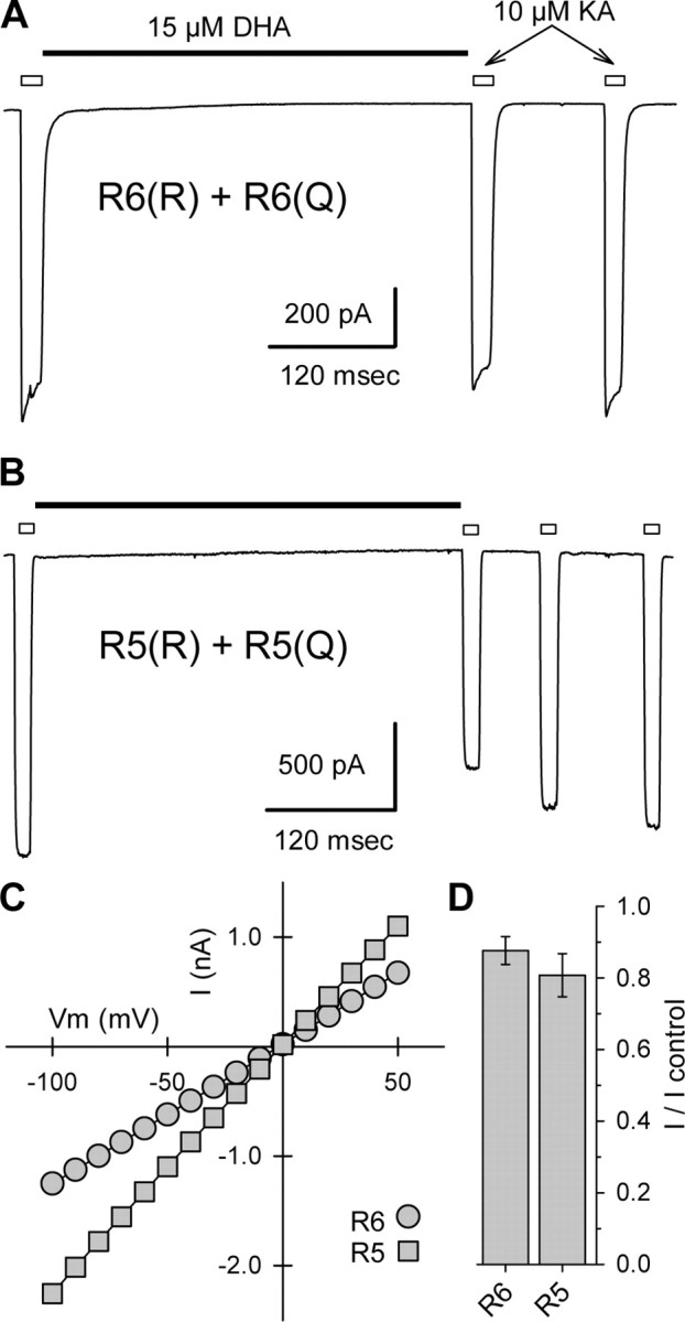
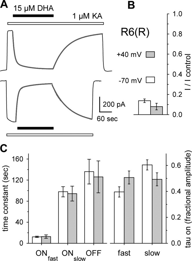
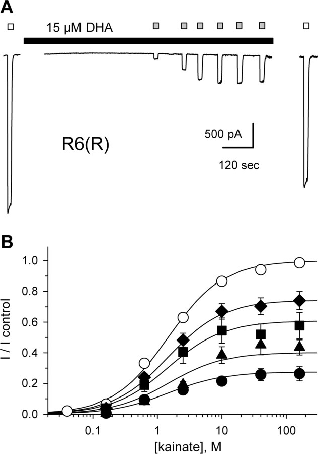
RNA editing within the pore loop controls the pharmacology and permeation properties of ion channels formed by neuronal AMPA and kainate receptor subunits. Genomic sequences for the glutamate receptor 2 (GluR2) subunit of AMPA receptors and the GluR5 and GluR6 subunits of kainate receptors all encode a neutral glutamine (Q) residue within the channel pore that can be converted by RNA editing to a positively charged arginine (R). Receptors comprised of unedited subunits are permeable to calcium and display inwardly rectifying current-voltage relationships, because of blocking of outward current by intracellular polyamines. In contrast, receptors that include edited subunits conduct less calcium, resist polyamine block, and have relatively linear current-voltage relationships. We showed previously that cis-unsaturated fatty acids, including arachidonic acid and docosahexanoic acid, exert a potent block of native kainate receptors as well as homomeric recombinant receptors formed by transfection of heterologous cells with cDNA for the GluR6(R) subunit. Here, we show that fatty acid blockade of recombinant homomeric and heteromeric kainate receptors is strongly dependent on editing at the Q/R site. Recombinant channels that include unedited subunits exhibit significantly weaker block than channels made up of fully edited subunits. Inhibition of fully edited channels is equivalent at voltages from -70 to +40 mV and is noncompetitive, consistent with allosteric regulation of channel function.


Figures

Figure 1.
Figure 1.
Differential block of homomeric GluR6(R) and GluR5(Q). A, B, HEK 293 cells transfected with GluR6(R) (A) or GluR5-2a(Q) (B) were tested with 300 μm kainate (KA) before (left) or 10 μm kainate after (right) treatment with Con A. Left, Superimposed whole-cell currents evoked by rapid application of kainate, indicated by the open bar, before and after exposure to 15 μm DHA. Right, Current evoked by kainate applications indicated by the open bars, before and after exposure to 15 μm DHA, as indicated by the solid bar. C, Inhibition as a function of DHA concentration. Current evoked by kainate (mean ± SEM; n = 5-15 cells per point) immediately after exposure to DHA is plotted as a fraction of the current before DHA. Smooth curves are the best fit of the following: I/I control = 0.1 + (0.9/(1 + ([DHA]/IC50))), where IC50 is the DHA concentration producing half-maximal inhibition, and n is the slope factor. R6(R) without Con A (○), IC50 = 136 nm, n = 1.4; with Con A (•), IC50 = 2.6 μm, n = 0.94. R5(Q) without Con A(□) and after Con A treatment (▪), IC50 = 55 μm; n = 1.1.
Figure 2.
Figure 2.
Homomeric GluR6(Q) resists inhibition. A, Current evoked by kainate applications indicated by open bars, before and after exposure to 15 μm DHA, as indicated by the solid bar. B, Normalized currents as a function of holding potential for cells transfected with R6(R) (n = 8 cells) or R6(Q) (n = 4 cells). C, Superimposed whole-cell currents evoked by rapid application of 300 μm kainate, indicated by the open bar, before and after exposure to 15 μm DHA. D, Current evoked by 1 or 10 μm kainate immediately after exposure to 15 μm DHA as a fraction of control current before DHA (3-9 cells per construct).
Figure 3.
Figure 3.
Strong block of heteromeric GluR5(R)/GluR6(R). A, Current evoked by 10 μm kainate (KA) applications indicated by open bars, before and after exposure to 15 μm DHA, as indicated by the solid bar. B, Current evoked by alternating applications of 10 μm kainate (open bars), and 10 μm ATPA (gray bars), before and after exposure to 15 μm DHA. C, Current evoked by 10 μm kainate (n = 9 cells) or 10 μm ATPA (n = 7 cells) immediately after exposure to 15 μm DHA as a fraction of control current before DHA. D, Normalized currents evoked by 10 μm kainate (n = 4 cells) or 10 μm ATPA (n = 7 cells) as a function of holding potential for cells transfected with R6(R) plus R5(R).
Figure 4.
Figure 4.
Weak inhibition of Q/R heteromeric receptors. A, B, Current evoked by 10 μm kainate applications indicated by open bars, before and after exposure to 15 μm DHA, as indicated by the solid bar. C, Normalized currents as a function of holding potential for cells transfected with R6(R) (n = 8 cells), R6(R) plus R5(R) (n = 4 cells), R5(Q) (n = 4 cells), R6(R) plus KA2 (n = 5 cells), R5(R) plus KA2 (n = 6 cells), R6(R) plus R5(Q) (n = 8 cells), or R6(Q) plus R5(R) (n = 6 cells). D, Inhibition as a function of DHA concentration. Current evoked by 10 μm kainate (n = 3-9 cells per point) immediately after exposure to DHA is plotted as a fraction of the current before DHA. Smooth curves are the best fit of the following: I/I control = 0.1 + (0.9/(1 + ([DHA]/IC50))), where IC50 is the DHA concentration producing half-maximal inhibition, and n is the slope factor. R6(R) plus R5(R), IC50 = 2.6 μm, n = 1.02; R6(R) plus KA2, IC50 = 36.4 μm, n = 2.73; R5(R) plus KA2, IC50 = 51.5 μm, n = 2.24; R6(R) plus R5(Q), IC50 = 86.1 μm, n = 1.01; R6(Q) plus R5(R), IC50 = 192.3 μm, n = 0.74. Data for R6(R) and R5(Q) are replotted from Figure 1C. Data from cultured rat hippocampal neurons (hippocampus, dark gray circles) were adapted from Wilding et al. (1998).
Figure 5.
Figure 5.
Weak inhibition of GluR6(Q/R) and GluR5(Q/R) receptors. A, B, Current evoked by 10 μm kainate applications indicated by open bars, before and after exposure to 15 μm DHA, as indicated by the solid bar. C, Currents plotted as a function of holding potential for the two cells shown in A and B. D, Current evoked by 10 μm kainate immediately after exposure to 15 μm DHA as a fraction of control current before DHA (n = 6 cells for each subunit combination).
Figure 6.
Figure 6.
Voltage-independent inhibition of R6(R). A, Currents evoked by 1 μm kainate (open bar) at -70 and +40 mV were strongly blocked by 15 μm DHA (solid bar). Superimposed lines show the best fits of the sum of two exponentials plus a constant to block onset or one exponential plus a constant to recovery from block. B, Steady-state block by 15 μm DHA at -70 mV (open bar; n = 18 cells) and +40 mV (shaded bar; n = 14 cells). C, Time constants (left) and relative amplitudes (right) for exponential fits to onset of block and recovery from block. Error bars represent SEM.
Figure 7.
Figure 7.
Noncompetitive inhibition of R6(R). A, Currents evoked by 10 mm kainate (open bars) in control solution or by 160 nm through 160 μm kainate (shaded bars) during exposure to 15 μm DHA (solid bar). B, Current (mean ± SEM; n = 6-14 cells per point) as a faction of the control response to 10 mm kainate versus kainate concentration. R6(R) in control solution (○) or during exposure to 0.3 μm (♦), 1 μm (▪), 5 μm (▴), or 15 μm (•) DHA. Smooth curves are the best fit of all data points to Equation 2. Error bars represent SEM.

References

    1. Bähring R, Bowie D, Benveniste M, Mayer ML (1997) Permeation and block of rat GluR6 glutamate receptor channels by internal and external polyamines. J Physiol (Lond) 502: 575-589. - PMC - PubMed
    1. Bernard A, Khrestchatisky M (1994) Assessing the extent of RNA editing in the TMII regions of GluR5 and GluR6 kainate receptors during rat brain development. J Neurochem 62: 2057-2060. - PubMed
    1. Bettler B, Boulter J, Hermans-Borgmeyer I, O'Shea-Greenfield A, Deneris E, Moll C, Borgmeyer U, Hollmann M, Heinemann S (1990) Cloning of a novel glutamate receptor subunit, GluR5: expression in the nervous system during development. Neuron 5: 583-595. - PubMed
    1. Bortolotto ZA, Clarke VR, Delany CM, Parry MC, Smolders I, Vignes M, Ho KH, Miu P, Brinton BT, Fantaske R, Ogden A, Gates M, Ornstein PL, Lodge D, Bleakman D, Collingridge GL (1999) Kainate receptors are involved in synaptic plasticity. Nature 402: 297-301. - PubMed
    1. Bowie D, Mayer ML (1995) Inward rectification of both AMPA and kainate subtype glutamate receptors generated by polyamine-mediated ion channel block. Neuron 15: 453-462. - PubMed

Publication types

LinkOut - more resources