Comparative Study
. 2005 Oct 12;25(41):9479-87.
doi: 10.1523/JNEUROSCI.0741-05.2005.

Neuronal basis of covert spatial attention in the frontal eye field

Affiliations
Comparative Study

Neuronal basis of covert spatial attention in the frontal eye field

Kirk G Thompson et al. J Neurosci. .

Abstract

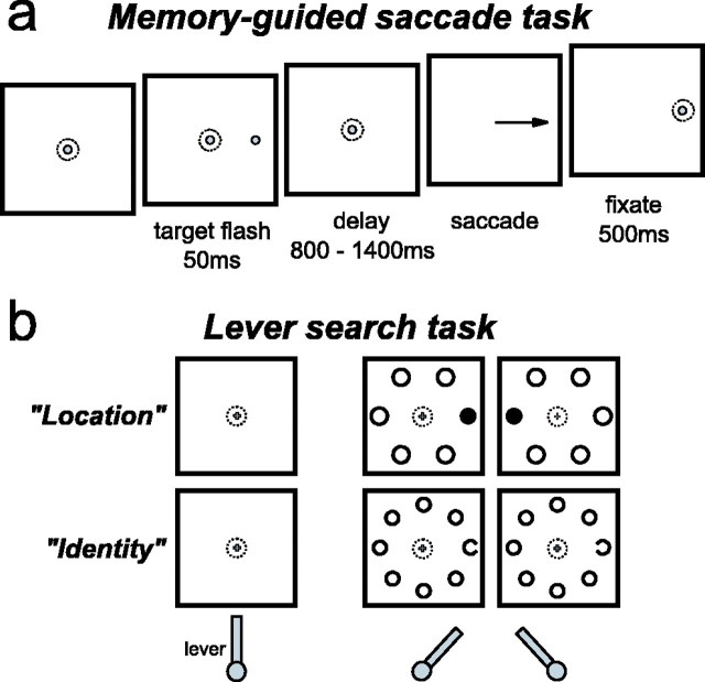
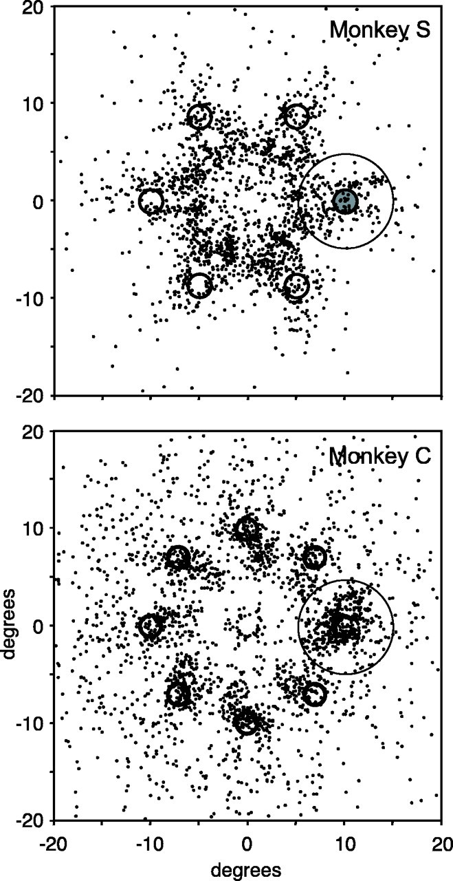
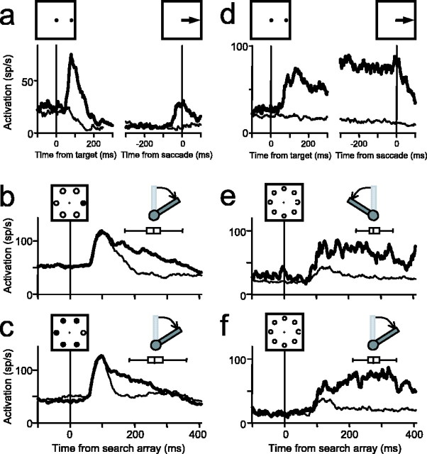
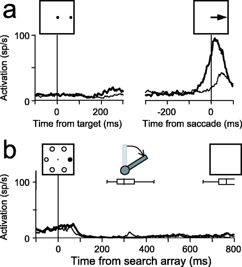
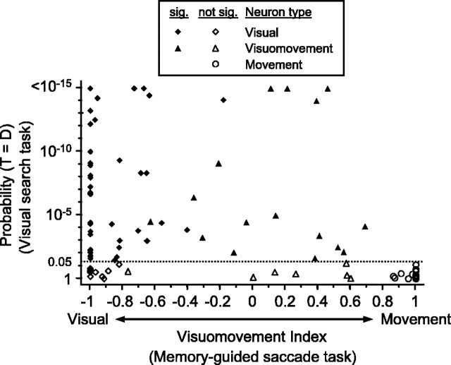
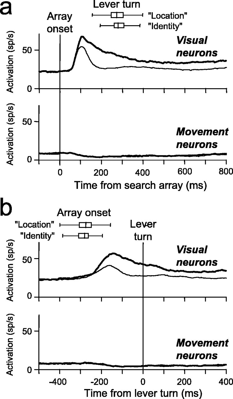
The influential "premotor theory of attention" proposes that developing oculomotor commands mediate covert visual spatial attention. A likely source of this attentional bias is the frontal eye field (FEF), an area of the frontal cortex involved in converting visual information into saccade commands. We investigated the link between FEF activity and covert spatial attention by recording from FEF visual and saccade-related neurons in monkeys performing covert visual search tasks without eye movements. Here we show that the source of attention signals in the FEF is enhanced activity of visually responsive neurons. At the time attention is allocated to the visual search target, nonvisually responsive saccade-related movement neurons are inhibited. Therefore, in the FEF, spatial attention signals are independent of explicit saccade command signals. We propose that spatially selective activity in FEF visually responsive neurons corresponds to the mental spotlight of attention via modulation of ongoing visual processing.


Figures

Figure 1.
Figure 1.
The tasks. a, The memory-guided saccade task. After the monkey fixated on a central spot, a peripheral stimulus identical to the fixation spot was flashed for 50 ms at one of six or eight locations. After a delay, the fixation spot was removed, and the monkey was instructed to make a saccade to the remembered target location. b, The manual lever search task. After the monkey grasped a lever in the vertical position, a small fixation cross appeared. After the monkey fixated on the central cross, a search array appeared in which one of the stimuli was different. In the location search (“Location”), the monkey was rewarded for turning the lever in the same direction as a different-colored stimulus in relation to the fixation cross. In the identity search (“Identity”), the monkey was rewarded for turning the lever in the same direction as the gap in the C stimulus.
Figure 2.
Figure 2.
Saccade behavior of monkey S during the location search task (top) and monkey C during the identity search task (bottom). Each point plots the endpoint of the first postreward saccade on each trial in which a saccade was made within a time window ending 500 ms after the search array was removed. The oddball stimulus is shown at the right horizontal position at 10° eccentricity, and the saccade endpoints were rotated and scaled accordingly for display. The circle around the target represents the 5° window in which saccades were counted as being made to the target location after the reward. These trials were removed from the neural activity analysis. For monkey S, a saccade was made after 1460 of 4710 trials (31%) and landed within 5° of the target on 188 trials (4%). For monkey C, a saccade was made after 2384 of 4966 trials (48%) and landed within 5° of the target on 394 trials (8%).
Figure 3.
Figure 3.
Representative examples of two visually responsive FEF neurons. a-c, The activity of a visuomovement neuron recorded during the memory-guided saccade task aligned on both target onset and saccade initiation (a) and the two complements of the location search task in which the target was red and the distractors were green (b) or in which the target was green and distractors were red (c). The monkey was rewarded for maintaining fixation on the central cross and indicating the location of the singleton target with a lever turn. d-f, The activity of a visual neuron recorded during the memory-guided saccade task aligned on both target onset and saccade initiation (d) and the identity search task in which the monkey was rewarded for indicating the direction of the C target among O distractors as pointing left (e) or right (f). For all plots, the activity on trials in which the target landed in the receptive field of the neurons (thick line) is plotted with the activity on trials in which no stimulus (a, d, thin line) or in which distractors (b, c, e, f, thin line) landed in the receptive field of the neurons. The box-whisker plot in each search panel indicates the median, quartiles, and range of lever turn reaction times. Diagrams showing the correct direction of the lever turn when the target was in the receptive field of the neurons are above each box-whisker plot.
Figure 4.
Figure 4.
A representative example of a movement neuron recorded during the memory-guided saccade task (a) and the location search task (b). Conventions are the same as in Figure 2, with the exception that the activity recorded during the lever search task is plotted until the time of the removal of the search array stimuli, which occurred ∼700 ms after search array presentation (b). The box-whisker plot in b indicates the median, quartiles, and range of lever turn reaction times. A diagram showing the correct direction of the lever turn when the target was in the receptive field of the neurons is above the box-whisker plot.
Figure 5.
Figure 5.
Statistical analysis of spatially selective activity in the covert visual search task as a function of neuron classification. The probability that the activity from 100 to 250 ms after search array presentation is the same on trials in which the target (T) is in the response field and on trials in which distractors (D) are in the response field is plotted as a function of the visuomovement index for each neuron. The visuomovement index is calculated as a contrast ratio of the visual and saccade-related responses recorded during the memory-guided saccade task. Neurons with values near -1 are dominated by a visual response, and neurons near +1 are dominated by saccade-related activity. Values near 0 indicate nearly equivalent visual and saccade-related activation. Visual neurons (diamonds) exhibit significant (sig.) visual responses and a no-movement response. Visuomovement neurons (triangles) have significant visual and movement responses. Movement neurons (circles) have no visual response and significant movement responses. Filled symbols indicate neurons with significantly different activity for the target and distractors in the covert visual search task. The horizontal dotted line indicates the probability threshold for a significant difference (p < 0.05). Six neurons (3 visual and 3 visuomovement) showed p < 10-15.
Figure 6.
Figure 6.
Pooled average activity from FEF neurons recorded during the lever search tasks aligned on the time of the search array presentation (a) and the time of the initiation of the lever turn (b). The activity of target-selective visually responsive neurons and movement neurons is shown separately. Thick lines plot the average activity on trials in which the target landed in the response field. Thin lines plot the average activity on trials in which only distractors landed in the response field. The spatial extent of the response field was based on activity recorded during the memory-guided saccade task (see Figs. 3a,d, 4a). For the movement neurons, the target-related and distractor-related activity was nearly identical and cannot be differentiated in the plots. The box-whisker plots show the median, quartile, and ranges of the lever turn reaction times (a) and search array presentation times (b) separately for the location and identity search tasks.

Comment in

References

    1. Astafiev SV, Shulman GL, Stanley CM, Snyder AZ, Van Essen DC, Corbetta M (2003) Functional organization of human intraparietal and frontal cortex for attending, looking, and pointing. J Neurosci 23: 4689-4699. - PMC - PubMed
    1. Beauchamp MS, Petit L, Ellmore TM, Ingeholm J, Haxby JV (2001) A parametric fMRI study of overt and covert shifts of visuospatial attention. NeuroImage 14: 310-321. - PubMed
    1. Bichot NP, Schall JD, Thompson KG (1996) Visual feature selectivity in frontal eye fields induced by experience in mature macaques. Nature 381: 697-699. - PubMed
    1. Bichot NP, Thompson KG, Chenchal Rao S, Schall JD (2001a) Reliability of macaque frontal eye field neurons signaling saccade targets during visual search. J Neurosci 21: 713-725. - PMC - PubMed
    1. Bichot NP, Chenchal Rao S, Schall JD (2001b) Continuous processing in macaque frontal cortex during visual search. Neuropsychologia 39: 972-982. - PubMed

Publication types