Comparative Study
. 2005 Oct 12;25(41):9544-53.
doi: 10.1523/JNEUROSCI.1826-05.2005.

Intracellular trafficking of histone deacetylase 4 regulates neuronal cell death

Affiliations
Comparative Study

Intracellular trafficking of histone deacetylase 4 regulates neuronal cell death

Timothy A Bolger et al. J Neurosci. .

Abstract

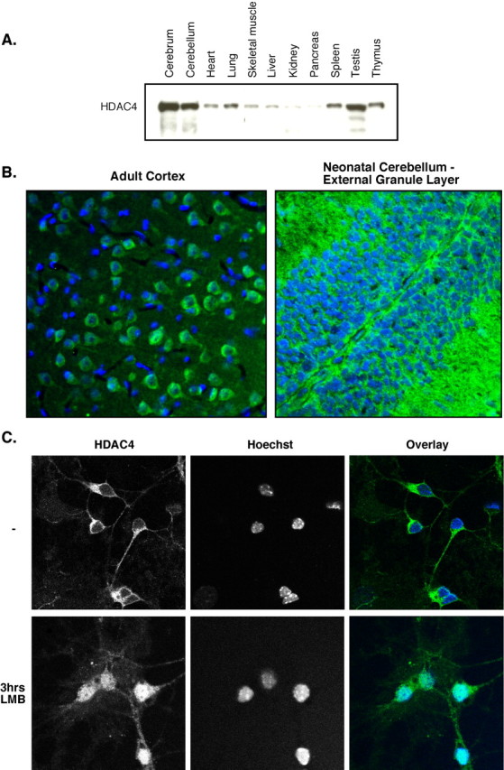
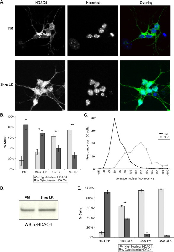
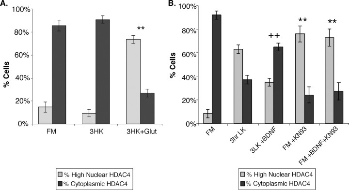
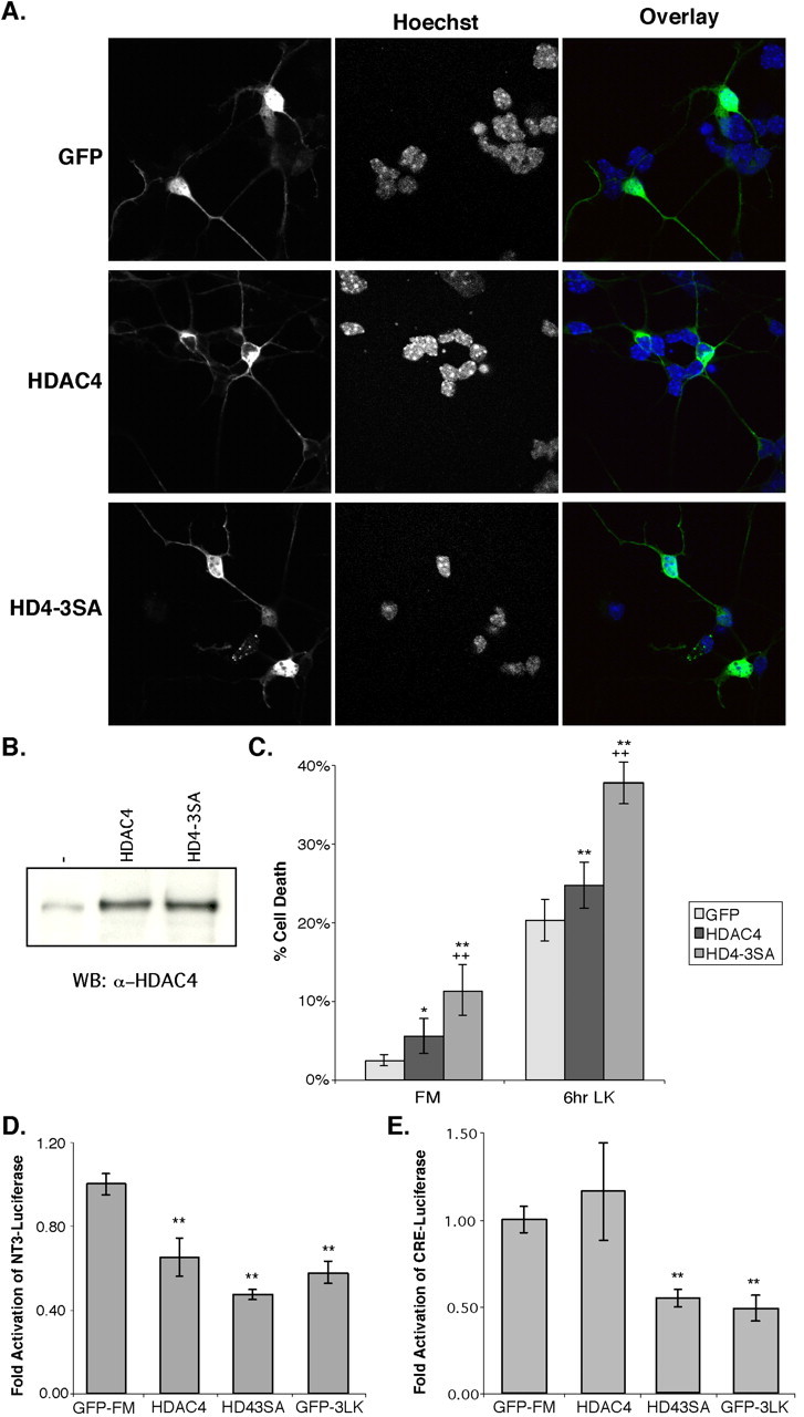
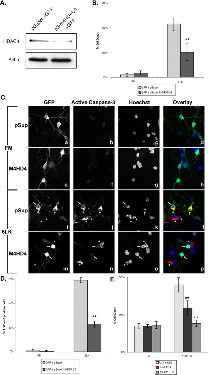
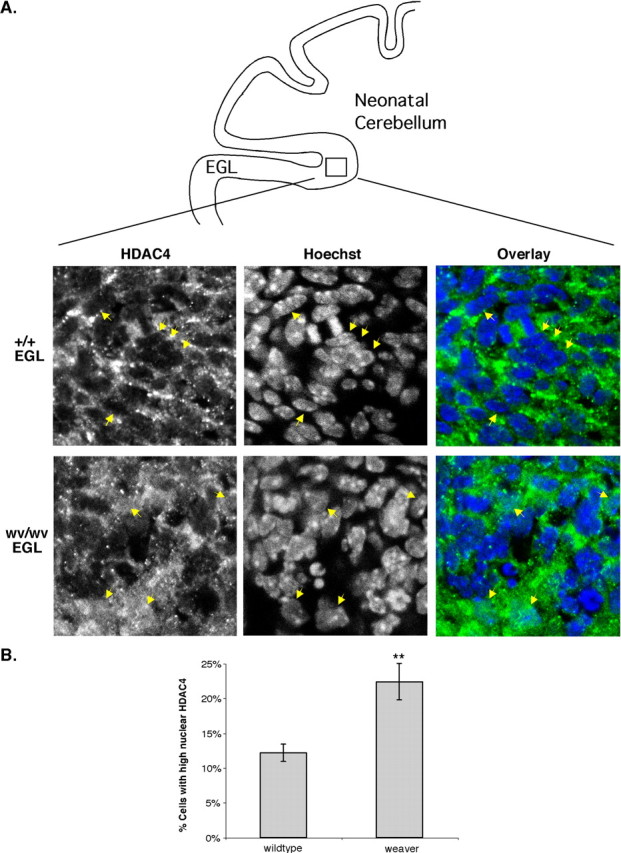
Histone deacetylase 4 (HDAC4) undergoes signal-dependent shuttling between the cytoplasm and nucleus, which is regulated in part by calcium/calmodulin-dependent kinase (CaMK)-mediated phosphorylation. Here, we report that HDAC4 intracellular trafficking is important in regulating neuronal cell death. HDAC4 is normally localized to the cytoplasm in brain tissue and cultured cerebellar granule neurons (CGNs). However, in response to low-potassium or excitotoxic glutamate conditions that induce neuronal cell death, HDAC4 rapidly translocates into the nucleus of cultured CGNs. Treatment with the neuronal survival factor BDNF suppresses HDAC4 nuclear translocation, whereas a proapoptotic CaMK inhibitor stimulates HDAC4 nuclear accumulation. Moreover, ectopic expression of nuclear-localized HDAC4 promotes neuronal apoptosis and represses the transcriptional activities of myocyte enhancer factor 2 and cAMP response element-binding protein, survival factors in neurons. In contrast, inactivation of HDAC4 by small interfering RNA or HDAC inhibitors suppresses neuronal cell death. Finally, an increase of nuclear HDAC4 in granule neurons is also observed in weaver mice, which harbor a mutation that promotes CGN apoptosis. Our data identify HDAC4 and its intracellular trafficking as key effectors of multiple pathways that regulate neuronal cell death.


Figures

Figure 1.
Figure 1.
HDAC4 is expressed highly in the brain and is primarily cytoplasmic in neurons. A, C57BL/6 adult male mice were dissected, and organs were homogenized. Fifty micrograms of each sample were run on SDS-PAGE and blotted with ab186 (HDAC4). B, Adult rat neocortex (left) and neonatal mouse cerebellum (right; EGL pictured) were immunofluorescently stained with ab186 (green) and Hoechst (blue). C, CGNs were isolated and cultured for 6 d in vitro in full media. Leptomycin B (LMB) was then added to the cells or not, and cells were stained after 3 h of treatment with ab186 and Hoechst dye. Representative images are shown.
Figure 2.
Figure 2.
HDAC4 translocates to the nucleus under conditions that induce cell death. After 5-7 d invitro, CGNs were left in FM or replaced with LK media. Cells were stained with ab186 and Hoechst dye. Representative images are shown in A. B, A quantification of the data is shown. Cells were considered cytoplasmic if cytoplasmic staining was brighter than nuclear staining. If the nuclear staining was equal to or greater than the cytoplasmic staining, the cell was counted as high-nuclear HDAC4. *p < 0.05; **p < 0.005 compared with FM. Three or more experiments were performed for each condition, and > 200 cells were counted in each experiment. C, After 5 d in vitro, CGNs were left in FM media or replaced with LK media for 3 h. Cells were then stained with ab186 and Hoechst. Images were taken of ∼120 cells in both FM and LK cells in each of two independent experiments, and average nuclear fluorescence of each cell was obtained and normalized. The histogram was then plotted of nuclear fluorescence versus number of cells for the FM and 3 h in LK conditions (3LK). The x-axis groups the cells by average nuclear fluorescence; for example, the “45” group represents all the cells with nuclear fluorescences between 30 and 45 units (on a 0-255 scale). The y-axis gives the number of cells in each group per 100 cells counted. D, CGN lysates from cells in FM and in LK media for 3 h were run on SDS-PAGE and immunoblotted with ab186. WB, Western blot. E, CGNs were transfected with GFP, HDAC4 (HD4), or HDAC4-3SA (3SA) immediately after isolation. After 2 d in vitro, the localization of HDAC4- and HD4-3SA-transfected CGNs was assessed in FM media and LK media after 3 h (3LK). Cells were stained with ab186 and Hoechst dye. **p < 0.005 versus HDAC4 in FM. Three or more experiments were performed for each condition, and > 100 cells were counted in each experiment. Error bars represent one SD above and below the mean.
Figure 3.
Figure 3.
Glutamate excitotoxicity, BDNF, and CaMK can regulate HDAC4 translocation in CGNs. A, After 6-7 d in vitro, CGNs were left in FM or replaced with no-serum HK media (3HK). One hundred micromolar l-glutamic acid (Glut) was then added to some HK samples to induce an excitotoxic response, and all cells were incubated for 3 h. Cells were then stained with ab186 and Hoechst dye. A quantification of the data are shown. **p < 0.001 compared with untreated HK. Three or more experiments were performed for each condition, and > 200 cells were counted in each experiment. B, After 2 d in vitro, CGNs were left in FM or replaced with LK media. Some cells (FM and LK) were treated with 100 ng/ml BDNF and/or 8 μm KN-93 at the same time as potassium deprivation (3 h). Cells were stained with ab186 and Hoechst dye. A quantification of the data is shown. ++p < 0.001 compared with untreated LK for 3 h; ** p < 0.001 compared with untreated FM. Three or more experiments were performed for each condition, and > 200 cells were counted in each experiment. Error bars represent one SD above and below the mean.
Figure 4.
Figure 4.
Overexpression of HDAC4 causes increased sensitivity to cell death and represses MEF2- and CREB-dependent activity. Immediately after isolation, CGNs were transfected with GFP, HDAC4, or HDAC4-3SA. After 2 d in vitro, the cells were left in FM or replaced with LK media for either 3 or 6 h. Cells were stained with Hoechst dye and either ab186 or αGFP. A, Representative images of cells in FM media are shown. B, Untransfected, HDAC4-transfected, or HD4-3SA-transfected CGNs were harvested and immunoblotted using ab186. WB, Western blot. C, Transfected cells were assessed for their viability by observing nuclei. Cells with nuclei that appeared condensed, fragmented, or perforated were considered dead/dying. *p < 0.05, **p < 0.01 compared with GFP control at each condition; ++p < 0.01 compared with wild-type HDAC4 at each condition. Five or more experiments were performed for each condition, and > 100 cells were counted in each experiment. D, CGNs were transfected with an NT-3(-1117)-luciferase fusion reporter (Shalizi et al., 2003) and GFP as a control, HDAC4, or HDAC4-3SA. After 2 d in vitro, some GFP cells were shifted to LK media for 3 h (GFP-3LK). Luciferase assays were then performed in triplicate, and the results were normalized to GFP in FM media for fold activation. A representative experiment is shown. **p < 0.01 compared with GFP control in FM. E, CGNs were transfected with a CRE-containing luciferase reporter (from the somatostatinpromoter) and GFP as a control, HDAC4, or HDAC4-3SA. After 2 d invitro, some GFP cells were shifted to LK media for 3 h (GFP-3LK). Luciferase assays were then performed in triplicate, and the results were normalized to GFP in FM media for fold activation. **p <0.001 versus CRE-luciferasein FM. A representative experiment is shown. HD4-3SA, HDAC4-3SA; 3 LK, 3 h in LK conditions. Error bars represent one SD above and below the mean.
Figure 5.
Figure 5.
Inhibition of HDAC4 by siRNA or pharmacological inhibitors reduces cell death of CGNs. Immediately after isolation, CGNs were cotransfected with GFP and either pSuper (pSup) or pSuper-m4HD4 (pS-m4HD4). A, After 2 d in vitro, cells were sorted for GFP positives by FACS. Lysates were equalized for cell number, then blotted for HDAC4 and actin. Band intensity was quantitated with NIH Image. B-D, Transfected cells were left in FM media (Ca-Ch) or switched to LK media for 6 h (Ci-Cp). The cells were stained with αGFP, Hoechst dye, and α-active-caspase-3 (C, D only). B, Nuclear morphology of GFP-positive cells was observed, and cells with nuclei that appeared condensed, fragmented, or perforated were considered dead/dying. **p <0.002 compared with control at 6LK. Four or more experiments were performed for each condition, and 100 or more cells were counted in each experiment. C, D, GFP-positive cells were assessed for the presence of positive staining for active caspase-3. Representative images are shown in C. Ci-Cp, Arrows point to transfected cells, and arrowheads to apoptotic untransfected cells. The data are quantitated in D. **p < 0.002 compared with control at 6LK. Three experiments were performed for eachcondition, and 200 or more cells were counted in each experiment. E, Untransfected CGNs after 6-8 d in vitro were pretreated with 1 μm TSA or 100 nm Trapoxin-A for 5 h or left untreated. Samples were then left in FM or replaced with LK media with TSA or TPX added for an additional 12 h. Cells were stained with Hoechst dye, and cell death was evaluated by examination of nuclei as above. **p < 0.002 compared with untreated LK media for 12 h (12 hr LK). Three or more experiments were performed for each condition, and > 400 cells were counted in each experiment. 6LK, 6 h in LK media. Error bars represent one SD above and below the mean.
Figure 6.
Figure 6.
Increased nuclear accumulation of HDAC4 is observed in weaver mutant mice. Weaver and wild-type cerebella from P7-P8 mice were sectioned and then stained with ab186 and Hoechst dye. A, Examples of the EGL of wild-type (+/+) and weaver homozygous (wv/wv) sections are shown. A schematic of a cross section of neonatal mouse cerebellum is included (top) to help orient the reader. In the wild-type section, arrows point to nuclear exclusion of HDAC4. In the weaver section, arrows point to nuclear accumulations of HDAC4. B, A quantification of HDAC4 localization in the sections is shown. Only cells in the EGL of the cerebellum were counted. If the nuclear staining was equal to or greater than the cytoplasmic staining, the cell was counted as high-nuclear HDAC4. Three or four animals were used of each genotype, and > 500 cells were counted per animal. **p < 0.001 compared with wild type. Error bars represent one SD above and below the mean.

References

    1. Benn SC, Woolf CJ (2004) Adult neuron survival strategies-slamming on the brakes. Nat Rev Neurosci 5: 686-700. - PubMed
    1. Bertos NR, Wang AH, Yang XJ (2001) Class II histone deacetylases: structure, function, and regulation. Biochem Cell Biol 79: 243-252. - PubMed
    1. Bonni A, Ginty DD, Dudek H, Greenberg ME (1995) Serine 133-phosphorylated CREB induces transcription via a cooperative mechanism that may confer specificity to neurotrophin signals. Mol Cell Neurosci 6: 168-183. - PubMed
    1. Boutillier AL, Trinh E, Loeffler JP (2003) Selective E2F-dependent gene transcription is controlled by histone deacetylase activity during neuronal apoptosis. J Neurochem 84: 814-828. - PubMed
    1. Chang S, McKinsey TA, Zhang CL, Richardson JA, Hill JA, Olson EN (2004) Histone deacetylases 5 and 9 govern responsiveness of the heart to a subset of stress signals and play redundant roles in heart development. Mol Cell Biol 24: 8467-8476. - PMC - PubMed

Publication types

LinkOut - more resources