. 2005 Oct 12;33(18):5728-39.
doi: 10.1093/nar/gki887. Print 2005.

The essential WD-repeat protein Rsa4p is required for rRNA processing and intra-nuclear transport of 60S ribosomal subunits

Affiliations

The essential WD-repeat protein Rsa4p is required for rRNA processing and intra-nuclear transport of 60S ribosomal subunits

Jesús de la Cruz et al. Nucleic Acids Res. .

Abstract

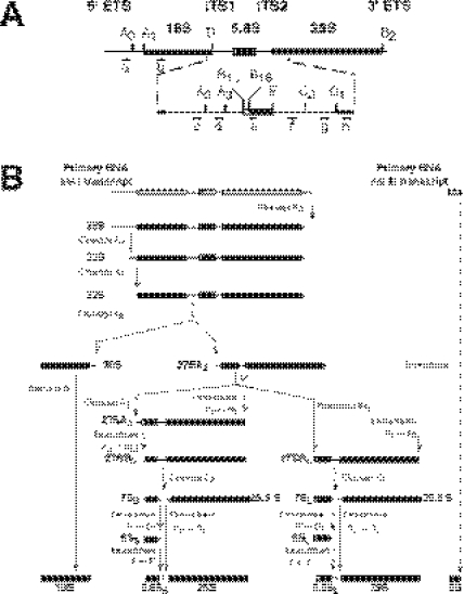
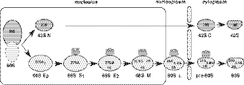
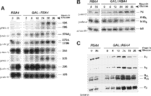
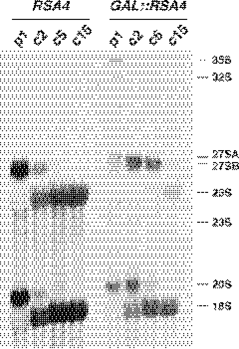
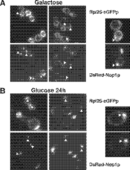
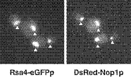
We report the characterization of a novel factor, Rsa4p (Ycr072cp), which is essential for the synthesis of 60S ribosomal subunits. Rsa4p is a conserved WD-repeat protein that seems to localize in the nucleolus. In vivo depletion of Rsa4p results in a deficit of 60S ribosomal subunits and the appearance of half-mer polysomes. Northern hybridization and primer extension analyses of pre-rRNA and mature rRNAs show that depletion of Rsa4p leads to the accumulation of the 27S, 25.5S and 7S pre-rRNAs, resulting in a reduction of the mature 25S and 5.8S rRNAs. Pulse-chase analyses of pre-rRNA processing reveal that, at least, this is due to a strong delay in the maturation of 27S pre-rRNA intermediates to mature 25S rRNA. Furthermore, depletion of Rsa4p inhibited the release of the pre-60S ribosomal particles from the nucleolus to the nucleoplasm, as judged by the predominantly nucleolar accumulation of the large subunit Rpl25-eGFP reporter construct. We propose that Rsa4p associates early with pre-60S ribosomal particles and provides a platform of interaction for correct processing of rRNA precursors and nucleolar release of 60S ribosomal subunits.


Figures

Figure 1
Figure 1
Pre-rRNA processing in S.cerevisiae. (A) Structure and processing sites of the 35S pre-rRNA. This precursor contains the sequences for the mature 18S, 5.8S and 25S rRNAs that are separated by two internal transcribed spacer sequences, ITS1 and ITS2, and flanked by two external transcribed spacer sequences, 5′ ETS and 3′ ETS. The mature rRNA species are shown as bars and the transcribed spacer sequences as lines. The processing sites and their locations as well as the various probes used are indicated. (B) Pre-rRNA processing pathway. The primary RNA pol I transcript undergoes covalent modifications (data not shown), and it is cleaved at its 3′ end to yield the 35S pre-rRNA, which is the longest detectable precursor. The 35S pre-rRNA is cleaved at site A0 to generate the 33S pre-rRNA. This molecule is subsequently processed at sites A1 and A2, resulting in the separation of the pre-rRNAs destined for the small and large ribosomal subunits. The final maturation of the 20S precursor takes place in the cytoplasm, where cleavage at site D yields the mature 18S rRNA. The 27SA2 precursor is processed by two alternative pathways that both lead to the formation of mature 5.8S and 25S rRNAs. In the major pathway, the 27SA2 precursor is first cleaved at site A3 and then, the 27SA3 precursor is exonucleolytically digested 5′→3′ up to site B1S to yield the 27SBS precursor. A minor pathway processes the 27SA2 or the 27SA3 molecule at site B1L by an as yet unknown mechanism, producing the 27SBL pre-rRNA. While processing at sites B1S and B1L is being completed, the 3′ end of the mature 25S rRNA is generated by 3′→5′ trimming to site B2. The subsequent processing of both 27SB species appears to be identical. Cleavage at site C2 generates the 25.5S and 7S pre-rRNAs. The 7S pre-rRNA is 3′→5′ trimmed to the 3′ end of the mature 5.8S rRNA. The 25.5S species is 5′→3′ digested to the mature 25S rRNA. The primary RNA pol III transcript is trimmed to the 3′ end of the mature 5S rRNA. The data presented in this study suggest that Rsa4p is required for efficient processing of all steps, leading from 27SA2 pre-rRNA to mature 25S and 5.8S rRNAs. For reviews on pre-rRNA processing and the known processing enzymes (4,5).
Figure 2
Figure 2
Current model for the formation, maturation and export of 66S pre-ribosomal particles in S.cerevisiae. A series of distinct particles are predicted to be intermediates during the synthesis of 60S r-subunits. These are termed, according to their position in the pathway, early 0 (E0), early 1 (E1), early 2 (E2) and middle (M) 66S pre-ribosomal particles and late (L) and cytoplasmic pre-60S ribosomal particles. All these particles are defined by the purification of complexes associated with TAP-tagged version of selected ribosomal subunit assembly factors (9,12,13,37,61). The pre-rRNAs abundantly associated with the different particles are indicated. Note that although in this figure the 5S rRNA assembles late in the pathway, its exact binding position is not clear. The data presented in this study and the literature suggest that Rsa4p associates with early pre-60S r-subunits and releases into the cytoplasm. Rsa4, grey barrel; nucleolus, rectangle; pre-ribosomal particles and mature r-subunits, light balloons; and nuclear envelope, rods. For further description of the ribosomal subunit assembly pathway (2,3,62).
Figure 3
Figure 3
RSA4 encodes a member of the WD-repeat protein family. Protein sequence is aligned to enhance the presence of the seven WD motifs. Those in bold face completely match the motif as it is found in Prosite (PDOC00574). The most highly conserved residues among the seven repeats are underlined. The hydrophilic region between domains IV and V is double underlined. In the N-terminal end, the putative nuclear location site is also underlined.
Figure 4
Figure 4
Conditional system for the phenotypic analysis. (A) Growth comparison of the strains WDG72 (GAL::RSA4) and W303-1A (RSA4). The cells were streaked on YPGal (Galactose) and YPD (Glucose) plates and incubated at 30°C for 3 days. (B) Growth curve of WDG72 at 30°C, after transferring exponential cells from YPGal to YPD medium, for up to 36 h. (C) Depletion of the Rsa4p protein; whole-cell extracts were prepared from W303-1A and WDG72, harvested at the indicated times after the shift to YPD medium. Equal amounts of protein were separated by 12% SDS–PAGE and Rsa4p was detected by western blotting using polyclonal rabbit antibodies.
Figure 5
Figure 5
Depletion of Rsa4p results in deficit in free 60S r-subunits and accumulation of half-mer polysomes. WDG72 (GAL::RSA4) was grown in YPGal (A) and then shifted to YPD for 24 h (B). Cells were harvested at an OD600 of ∼0.8 and equal amounts of cells extracts (10 A260 units) were resolved in 7–50% (w/v) sucrose gradients. The A254 was read continuously. Sedimentation is from left to right. The peaks of free 40S and 60S ribosomal subunits, 80S free couples/monosomes and polysomes are indicated. Half-mers are indicated by arrows.
Figure 6
Figure 6
Effects of Rsa4p depletion on steady-state levels of pre-rRNAs and mature rRNAs. The strains W303-1A (RSA4) and WDG72 (GAL::RSA4) were grown in YPGal medium and then shifted to YPD medium. Cells were harvested at indicated times and total RNAs were extracted. (A) Equal amounts of total RNA (5 µg) were resolved on a 1.2% agarose–formaldehyde gel and transferred onto a nylon membrane. The same membrane was hybridized with different probes. (B) Equal amounts of total RNA (2.5 µg) were resolved on a 7% polyacrylamide–urea gel, transferred onto a nylon membrane and hybridized consecutively with different probes. (C) Equal amounts of total RNA (5 µg) were used for primer extension analysis. Probe g was labelled and used for the reactions. Note that this probe allows detection of 27SA2 (as the stop at site A2), 27SA3 (as the stop at site A3), and both 27SB (as stops at sites B1L and B1S) and 25.5S (as the stop at site C2). Probe names are indicated between parentheses on the left (except for probe 5S, see Figure 1A for their location in the 35S pre-rRNA).
Figure 7
Figure 7
Depletion of Rsa4p inhibits pre-rRNA processing of 27S precursors. The strains W303-1A (RSA4) and WDG72 (GAL::RSA4) were grown in YPGal medium and then shifted for 24 h to SD-Met medium. Cells were pulse-labelled with [methyl-3H]methionine for 1 min, and then chased with a large excess of ice-cold methionine for 2, 5 and 15 min. Total RNA was extracted and 20 000 c.p.m. was loaded and separated on a 1.2% agarose–formaldehyde gel, transferred onto a nylon membrane and visualized by fluorography. The positions of the different pre-rRNAs and mature rRNAs are indicated.
Figure 8
Figure 8
Depletion of Rsa4p leads to accumulation of Rpl25p-eGFP in the nucleolus. WDG72 (GAL::RSA4) was transformed with Rpl25p-eGFP and DsRed-Nop1p plasmids. Selected candidates were grown in liquid SGal-Leu-Ade medium (galactose) (A) and then shifted to liquid SD-Leu-Ade medium (glucose) for 24 h (B). Cells were harvested, washed and resuspended in water and inspected in the fluorescence microscope. Selected cells are shown in a magnified picture. Triangles point to nucleolar fluorescence.
Figure 9
Figure 9
Rsa4p localized predominantly to the nucleolus. In vivo localization of Rsa4p-eGFP and DsRed-Nop1p was determined by fluorescence microscopy. Triangles point to nucleolar fluorescence.

References

    1. Olson M.O., Dundr M., Szebeni A. The nucleolus: an old factory with unexpected capabilities. Trends Cell Biol. 2000;10:189–196. - PubMed
    1. Fromont-Racine M., Senger B., Saveanu C., Fasiolo F. Ribosome assembly in eukaryotes. Gene. 2003;313:17–42. - PubMed
    1. de la Cruz J., Kressler D., Linder P. Ribosomal subunit assembly. In: Olson M.O.J., editor. Nucleolus. Georgetown: Kluwer academic. Landes Bioscience/eurekah.com; 2004. pp. 258–285.
    1. Venema J., Tollervey D. Ribosome synthesis in Saccharomyces cerevisiae. Annu. Rev. Genet. 1999;33:261–311. - PubMed
    1. Raué H.A. Pre-ribosomal RNA processing and assembly in Saccharomyces cerevisiae: the machine that makes the machine. In: Olson M.O.J., editor. Nucleolus. Georgetown: Kluwer academic. Landes Biosciences/Eurekah.com; 2004. pp. 199–222.

Publication types

MeSH terms