. 2005 Oct 12;33(18):5851-60.
doi: 10.1093/nar/gki898. Print 2005.

Guanine tetraplex topology of human telomere DNA is governed by the number of (TTAGGG) repeats

Affiliations

Guanine tetraplex topology of human telomere DNA is governed by the number of (TTAGGG) repeats

Michaela Vorlícková et al. Nucleic Acids Res. .

Abstract

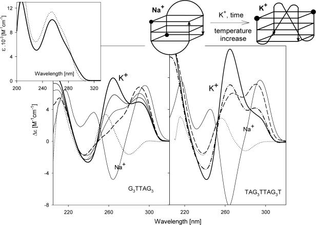
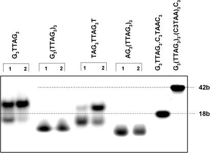
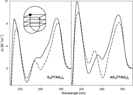
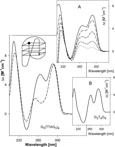
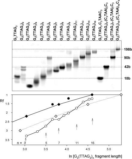
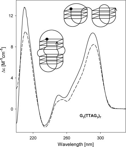
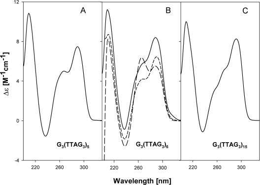
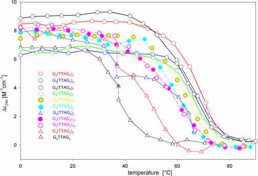
Secondary structures of the G-rich strand of human telomere DNA fragments G3(TTAG3)n, n = 1-16, have been studied by means of circular dichroism spectroscopy and PAGE, in solutions of physiological potassium cation concentrations. It has been found that folding of these fragments into tetraplexes as well as tetraplex thermostabilities and enthalpy values depend on the number of TTAG3 repeats. The suggested topologies include, e.g. antiparallel and parallel bimolecular tetraplexes, an intramolecular antiparallel tetraplex, a tetraplex consisting of three parallel chains and one antiparallel chain, a poorly stable parallel intramolecular tetraplex, and both parallel and antiparallel tetramolecular tetraplexes. G3(TTAG3)3 folds into a single, stable and very compact intramolecular antiparallel tetraplex. With an increasing repeat number, the fragment tetraplexes surprisingly are ever less thermostable and their migration and enthalpy decrease indicate increasing irregularities or domain splitting in their arrangements. Reduced stability and different topology of lengthy telomeric tails could contribute to the stepwise telomere shortening process.


Figures

Figure 1
Figure 1
CD spectra of G3TTAG3 (left) and TAG3TTAG3T (right) measured at 25°C (dots) in 1 mM Na phosphate plus 0.3 mM EDTA, pH 7 immediately after thermal denaturation. Measurements in 10 mM potassium phosphate plus 0.15 M KCl were carried out (left): immediately (dashes) and 1, 3 and 10 days (from the thinnest to the thickest line) after K+ addition. The samples were kept and measured at 25°C; (right): after two days keeping at 0°C and measured at 0°C (dashes) and 25°C (dash–dots) and after three days keeping at 25°C and measured at 25°C (thick line). The thin full line spectra in both panels correspond to the samples kept for two days in 10 mM Na phosphate plus 0.15 M NaCl at 0°C and measured at 0°C. Inset: UV absorption spectra of G3TTAG3 in 10 mM K phosphate plus 0.15 M KCl at 91°C (dots) and 25°C (solid line). The sketch shows the antiparallel and parallel bimolecular tetraplexes of TAG3TTAG3T. The balls in the sketch refer to the oligonucleotide 5′ ends.
Figure 2
Figure 2
Electrophoresis run in the Robinson–Britton buffer plus 0.15 M KCl, pH 7.2, at 2°C. The samples were incubated one day in the electrophoretic buffer at 0°C (lanes 1) and at 37°C (lanes 2). The sample of G3TTAG3 was loaded immediately (lane 1) on the gel and after two days incubation (lane 2) at room temperature. The heteroduplexes G3TTAG3•C3TAAC3 and G3(TTAG3)3•(C3TAA)3C3 were used as markers for mobility of 18 and 42 base long fragments, respectively.
Figure 3
Figure 3
CD spectra of G3(TTAG3)3 and AG3(TTAG3)3 in 1 mM Na phosphate plus 0.3 mM EDTA, pH 7, measured at 0°C (dashes) and in 10 mM K phosphate plus 0.15 M KCl (solid lines). The same spectra were obtained at 0°C and at room temperature. The sketch shows the tetraplex structure of G3(TTAG3)3 or AG3(TTAG3)3. The ball in the sketch refers to the oligonucleotide 5′ end.
Figure 4
Figure 4
CD spectra of G3(TTAG3)4 in 1 mM Na phosphate plus 0.3 mM EDTA, pH 7 (dashes) and in 10 mM K phosphate plus 0.15 M KCl (solid line) measured at 0°C. Inset A: CD spectra of G3(TTAG3)4 in 10 mM K phosphate plus 0.15 M KCl measured at 45, 61, 68, 73 and 79°C (from the solid to the dotted line). Inset B: CD spectrum of G3T4G4 tetraplex measured in the same solution at 0°C. The sketch shows a suggested structure of G3(TTAG3)4 tetraplex. The ball refers to the oligonucleotide 5′ end.
Figure 5
Figure 5
Upper panel: electrophoresis of the telomere fragments (primary structures given above the lanes) run in the Robinson-Britton buffer plus 0.15 M KCl, pH 7.2, at 2°C. The samples were incubated one day in the electrophoretic buffer before loading on the gel. The heteroduplexes with complementary C-rich strands serve as markers for 18, 42, 90 and 198 base long fragments. Bottom panel: Rf—mobility related to that of G3(TTAG3)16•(C3TAA)16C3 heteroduplex as a function of natural logarithm of fragment lengths: heteroduplexes serving as markers (diamonds); bimolecular tetraplexes (solid circles); intramolecular tetraplexes (open circles). The dotted line with open diamonds simulates mobility of single-stranded molecules.
Figure 6
Figure 6
CD spectra of G3(TTAG3)7 in 1 mM Na phosphate plus 0.3 mM EDTA, pH 7 (dashed line) and 10 mM K phosphate plus 0.15 M KCl (solid line), both at 0°C. The sketches show two alternative arrangements of the G3(TTAG3)7 tetraplex fitting the results obtained. The ball refers to the oligonucleotide 5′ end.
Figure 7
Figure 7
CD spectra of (A) G3(TTAG3)5, (B) G3(TTAG3)6 and (C) G3(TTAG3)15 in 10 mM potassium phosphate plus 0.15 M KCl measured at 0°C. In addition, the (B) contains CD spectra of G3(TTAG3)6 in the same solvent but measured at 37°C immediately after increasing temperature (short dashes), and after one day incubation at 37°C (long dashes).
Figure 8
Figure 8
Temperature dependences of the telomere DNA fragments in 10 mM potassium phosphate plus 0.15 M KCl, pH 7, monitored by changes in Δɛ290. All points, with the exception of those marked with a cross, correspond to equilibrium values. The equilibrium was reached within 110 min with G3TTAG3 and 20 h with G3(TTAG3)2 and G3(TTAG3)6. Solid lines and open circles: G3(TTAG3)3 (red), G3(TTAG3)4 (blue), G3(TTAG3)5 (green), and G3(TTAG3)7 (black); dashed lines and solid circles: G3(TTAG3)8 (yellow), G3(TTAG3)9 (cyan); G3(TTAG3)15 (violet solid circles) and G3(TTAG3)16 (violet open circles); thin solid lines and triangles: G3(TTAG3)2 (blue), G3(TTAG3)6 (red), and G3TTAG3(black).

Similar articles

Cited by

References

    1. Blackburn E.H., Greider C.W. Telomeres. Plainview, NY, USA: Cold Spring Harbor Laboratory; 1995.
    1. Cech T.R., Nakamura T.M., Lingner J. Telomerase is a true reverse transcriptase. A review. Biochemistry (Mosc) 1997;62:1202–1205. - PubMed
    1. Londono-Vallejo A., DerSarkissian H., Cazes L., Thomas G. Differences in telomere length between homologous chromosomes in humans. Nucleic Acids Res. 2001;29:3164–3171. - PMC - PubMed
    1. Henderson E., Hardin C.C., Walk S.K., Tinoco I.J., Blackburn E.H. Telomeric DNA oligonucleotides form novel intramolecular structures containing guanine–guanine base pairs. Cell. 1987;51:899–908. - PubMed
    1. Choi K.-H., Choi B.-S. Formation of a hairpin structure by telomere 3′ overhang. Biochim. Biophys. Acta. 1994;1217:341–344. - PubMed

Publication types