. 2005 Dec 15;288(2):582-94.
doi: 10.1016/j.ydbio.2005.09.024. Epub 2005 Oct 11.

Odd-skipped related 1 (Odd 1) is an essential regulator of heart and urogenital development

Affiliations

Odd-skipped related 1 (Odd 1) is an essential regulator of heart and urogenital development

Qingru Wang et al. Dev Biol. .

Abstract

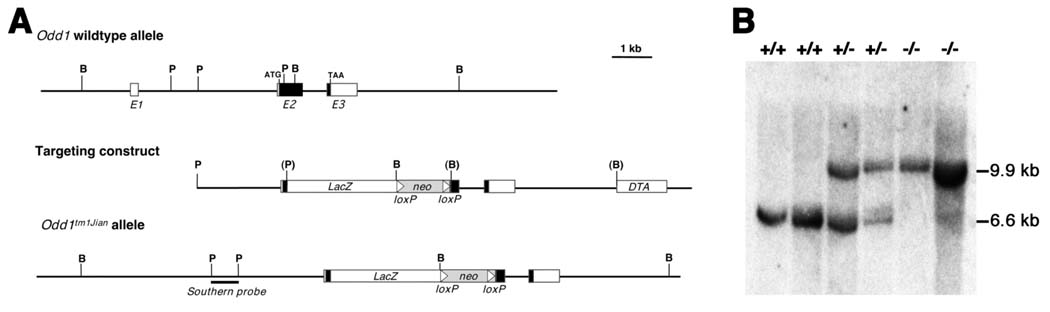
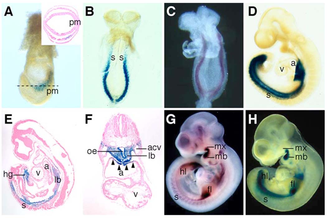
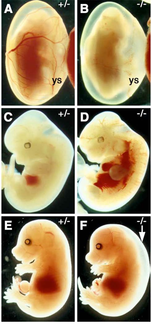
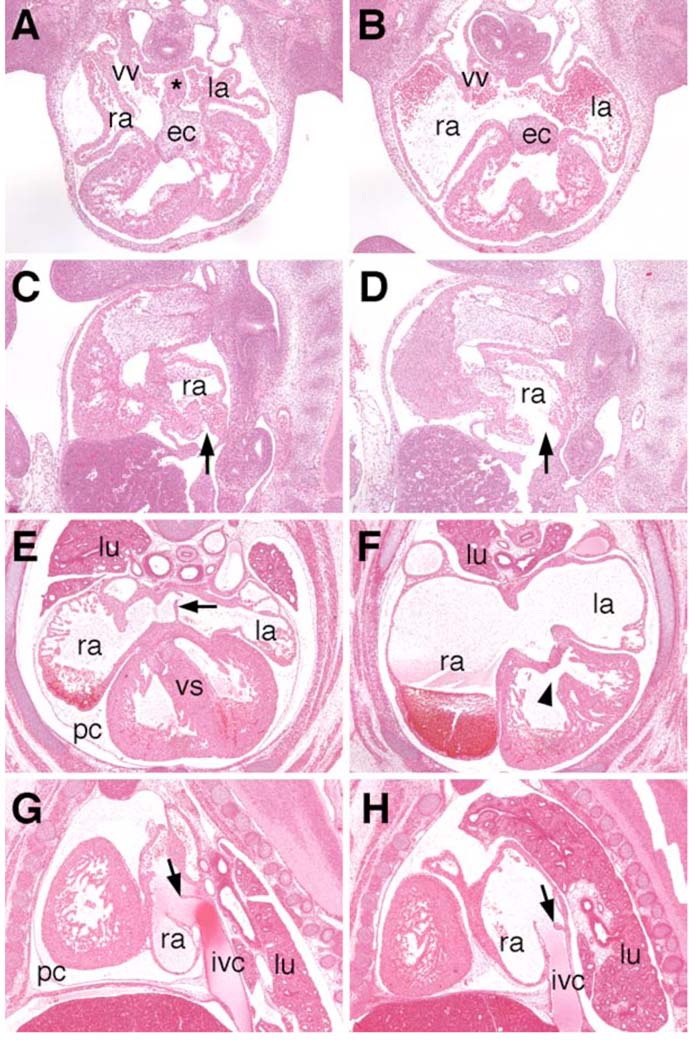
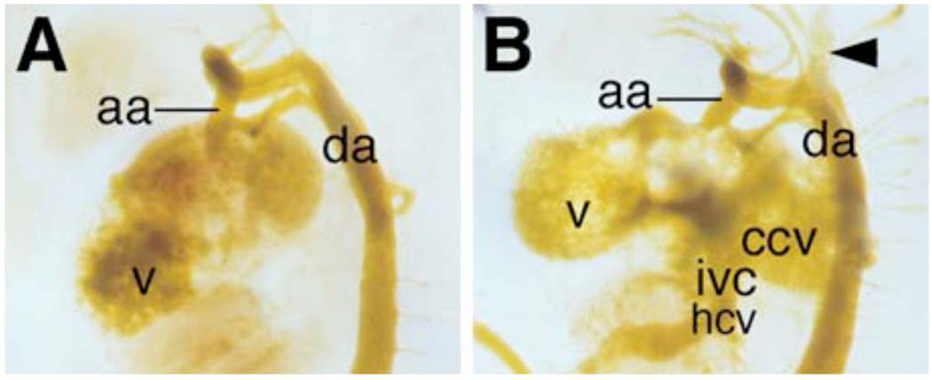
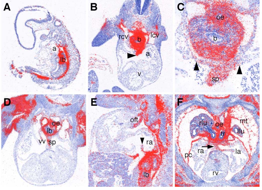
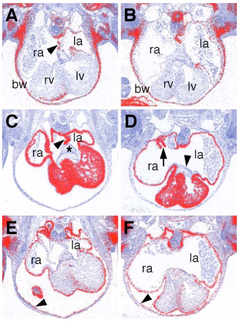
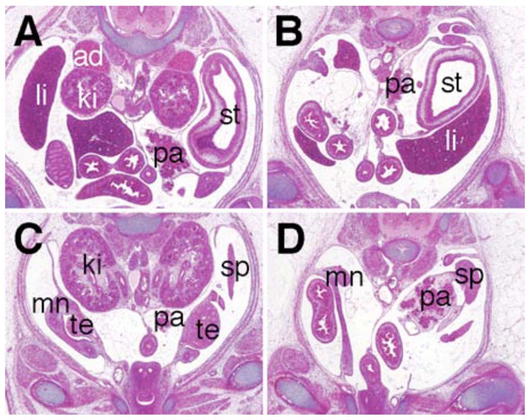
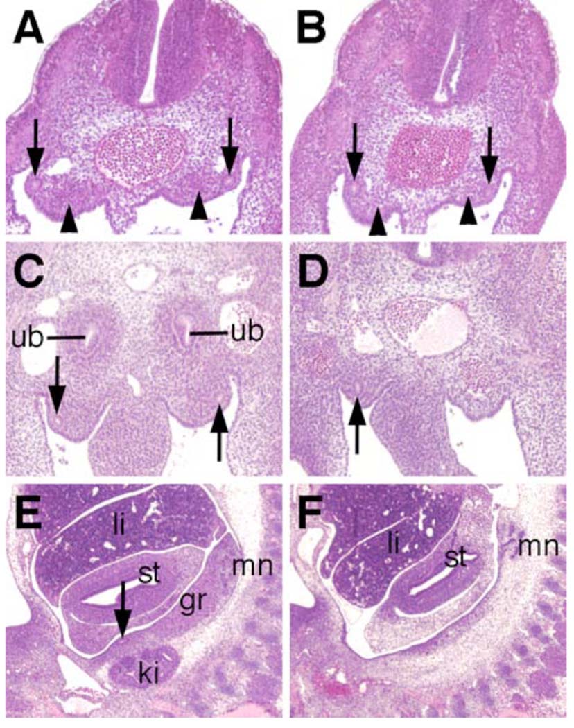
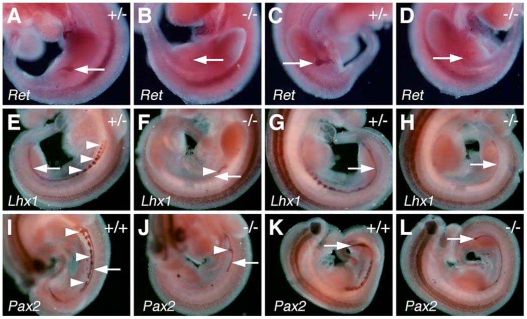
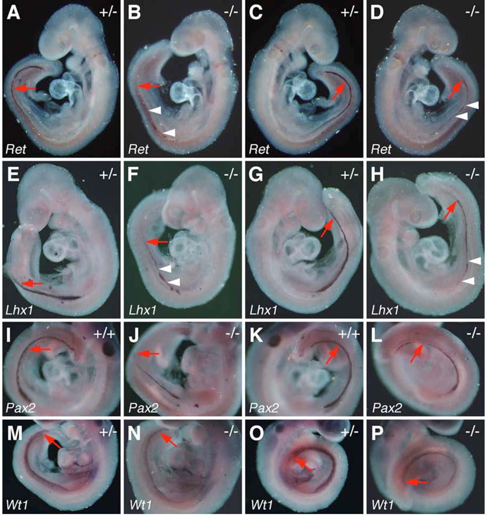
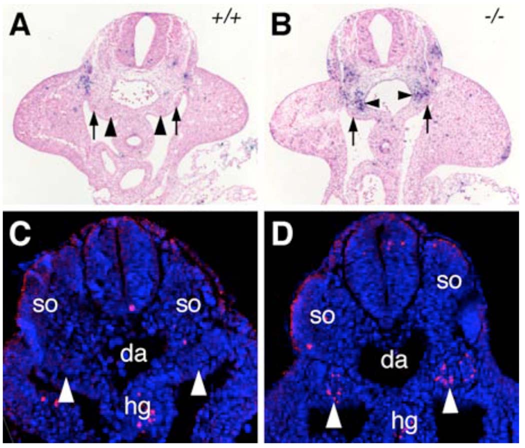
The Odd-skipped related 1 (Odd 1) gene encodes a zinc finger protein homologous to the Drosophila Odd-skipped class transcription factors that play critical roles in embryonic patterning and tissue morphogenesis. We have generated mice carrying a targeted null mutation in the Odd 1 gene and show that Odd 1 is essential for heart and intermediate mesoderm development. Odd 1(-/-) mutant mouse embryos fail to form atrial septum, display dilated atria with hypoplastic venous valves, and exhibit blood backflow from the heart into systemic veins. In contrast to other transcription factors implicated in atrial septum development, Odd 1 mRNA expression is restricted to the central dorsal domain of the atrial myocardium during normal heart development. Moreover, expression patterns of known key regulatory genes of atrial septum development, including Nkx2.5, Pitx2, and Tbx5, are unaltered in the developing heart in Odd 1(-/-) mutants compared to that of the wild-type littermates. Furthermore, homozygous Odd 1(-/-) mutant embryos exhibit complete agenesis of adrenal glands, metanephric kidneys, gonads, and defects in pericardium formation. Detailed molecular marker analyses show that key regulators of early intermediate mesoderm development, including Lhx1, Pax2, and Wt1, are all down-regulated and nephrogenic mesenchyme undergoes massive apoptosis, resulting in disruption of nephric duct elongation and failure of metanephric induction in the Odd 1(-/-) mutant embryos. These data provide new insights into the molecular mechanisms underlying heart morphogenesis and urogenital development.


Figures

Fig. 1
Fig. 1
Targeted disruption of the mouse Odd1 gene. (A) The Odd1 gene consists of three exons spanning approximately 6 kb of genomic DNA. Boxes indicate exons, with the protein-coding region marked in black. The positions of the translation start (ATG) and stop (TAA) codons are also indicated. Restriction sites are: B, BamHI; P, PstI. The targeting vector used the 2.1 kb PstI fragment containing the intron 1/exon 2 junction as the 5’ arm and the 5.3 kb BamHI fragment located 335 bp downstream as the 3’ arm. A modified bacterial lacZ gene and a loxP-flanked neo expression cassette were inserted in between the arms and a diptheria toxin A (DTA) expression cassette was cloned 3’ to the 3’ arm for negative selection against random integration. (B) Southern hybridization analysis of E11.5 embryonic DNA samples from a litter of F1 heterozygous intercross. The genomic DNA samples were digested with BamHI, separated by electrophoresis through a 0.8% agarose gel, transferred onto a Zetaprobe nylon memberane (BioRad), and hybridized with random prime-labeled probes made from the 700 bp PstI fragment isolated from the Odd1 genomic region 5’ to the targeted region. The 6.6 kb BamHI fragment corresponding to the wildtype allele was detected in wildtype and heterozygous embryos, while the 9.9 kb mutant allele-specific fragment was only detected in heterozygous and homozygous mutants. +/+, wildtype; +/−, heterozygous; −/−, homozygous mutant.
Fig. 2
Fig. 2
β-galactosidase fusion protein expression from the Odd1tm1Jian allele recapitulates Odd1 mRNA expression pattern during mouse embryogenesis. (A) Whole mount X-gal staining shows strong Odd1-LacZ expression (blue color) in a 7.5 dpc mouse embryo. The inset is a transverse section of a X-gal stained E7.5 embryo, which shows Odd1-LacZ expression in a few cells mixed with non-expressing cells in the intermediate region of the mesoderm layer. No staining is detected in the primitive streak cells. (B) At E8.5, Odd1-LacZ is expressed throughout the intermediate mesoderm caudal to the cardiac cresent. The expression pattern is identical to Odd1 mRNA expression pattern at this stage (C). (D) By E9.5, Odd1-LacZ expression is detected, in addition to the intermediate mesoderm, at the dorsal region of the atrium and in the caudal somites. (E,F) Sagittal and transverse sections, respectively of E9.5 heterozygous embryos stained with X-gal. Strong Odd1-LacZ expression is detected in the lung bud mesenchyme, the fore- and hindgut endoderm, and in the dorsal atrial wall. (G,H) By E10.5, both Odd1-LacZ (G) and Odd1 mRNA are expressed in identical patterns in the first branchial arches, limb buds, and somites. a, atrium; acv, anterior cardinal vein; fl, forelimb bud; hg, hindgut; hl, hindlimb bud; lb, lung bud; mb, mandibular process; mx, maxillary process; oe, oesophagous; pm, primitive streak; s, somite; v, ventricle.
Fig. 3
Fig. 3
Odd1 homozyogus mutant embryos die prenatally. (A, C, E) newly dissected Odd+/− and (B, D, F) Odd−/− mutant embryos. Most Odd1−/− mutant embryos die at E12.0 (B) with blood pooled in the heart and major veins (D). (F) An E15.5 Odd−/− mutant embryo showing generalized edema (arrow). ys, yolk sac.
Fig. 4
Fig. 4
Histological analyses of Odd1 mutant heart defects. (A, B) Transverse sections of the wild-type and Odd−/− mutant embryos at E11.5 showing absence of septum primum (marked by the asterisk in A) and dilated atria in the Odd−/− mutant. (C, D) Sagittal sections of Odd+/− (C) and Odd−/− mutant (D) embryos at E11.5 show absence of venous valve in the homozygous mutant. The entrance of the inferior vena cava is wide open in the homozygous mutant (arrow in D). (E, F) Transverse sections of Odd+/− (E) and Odd−/− mutant (F) embryos at E15.5. Arrow in E points to the primary atrial septum, which is absent in the Odd−/− mutant heart (F). The Odd−/− mutant (F) also had severely dilated atria, defective atrioventricular junction and ventricular septum (arrowhead), as well as lack of parietal pericardium surrounding the ventricles. (G,H) Sagittal sections of wildtype (G) and Odd−/− mutant (H) embryos show malformed venous valve at the entrance of the inferior vena cava. The left leaflet of the venous valve in the mutant heart is stumped and less than half the length of that in the control embryos (arrows in G and H). The lung tissues are clumped together in the homozygous mutants (F,H) compared to the well-extended left and right lungs in the wildtype (E) and heterozygous (G) embryos. ec, endocardial cushion; ivc, inferior vena cava; la, left atrium; lu, lung; pc, pericardium; ra, right atrium; vv, venous valve.
Fig. 5
Fig. 5
Odd1−/− mutant embryos suffer blood backflow from the heart into the inflow tract. (A) An E11.5 wildtype embryo showing normal blood flow from the heart into the outflow tract as traced by yellow casting dye. (B) An Odd−/− littermate of the embryo shown in A was similarly injected with yellow casting dye into the left ventricle. Although most of casting dye followed blood flow into the aortas, a significant amount of the dye backflowed into the common cardinal vein, the anterior cardinal vein (arrowhead), and hepato-cardiac vein. aa, asending aorta; ccv, common cardinal vein; da, dorsal aorta; hcv, hepato-cardiac vein; ivc, inferior vena cava; v, ventricle.
Fig. 6
Fig. 6
Odd1 mRNA expression during heart development. (A) Sagittal section of an E9.5 embryo showing Odd1 mRNA expression (red color) in the dorsal atrial wall. (B) Transverse section of an E10.5 embryo showing abundant Odd1 mRNA expression in the developing septum primum (arrowhead) and the dorsal atrial wall. Odd1 mRNA is also highly expressed in the cardinal vein. (C) Higher magnification of the lung bud region of the section in B. Odd1 mRNA is abundantly expressed in the oesophagus endoderm, the lung bud mesenchyme, the septum primum, and the dorsal atrial wall. Arrowheads mark the left and right boundaries of the dorsal atrial expression domain. (D, E) Transverse (D) and sagittal (E) sections of E11.5 embryos showing Odd1 mRNA expression in the left, but not right leaflet of the newly formed venous valves. (F) Frontal section through the dorsal thoracic region an E13.5 embryo showing strong expression of Odd1 mRNA in the dorsal atrial wall flanking the atrial septum (arrow) and in the mesothelium including the parietal pericardium. Low levels of Odd1 mRNA is detected in the lung mesenchyme at this stage. a, atrium; b, main bronchus; la, left atrium; lb, lung bud; lcv, left cardinal vein; llu, left lung; mt, mesothelium; oe, oesophagus; oft, outflow tract; pc, parietal pericardium; ra, right atrium; rcv, right cardinal vein; rv, right ventricle; v, ventricle; vv, venous valve; sp, septum primum; tr, trachea.
Fig. 7
Fig. 7
Comparison of molecular marker expression during heart development in the Odd1−/− mutant (B, D, F) and wildtype control (A, C, E) embryos at E11.5. (A, B) Pitx2 mRNA expression is restricted to the left atria in both wildtype (A) and Odd1−/− mutant embryos. Arrowhead in A points to the primary atrial septum, which is absent in the Odd1−/− mutant embryo. (C, D) Nkx2.5 mRNA is expressed throughout the myocardium in both the wildtype (C) and the mutant (D) embryos. Arrowhead in C points to the primary atrial septum, which is absent in the mutant embryos. The asterisk in C marks the mesodermal cap of the atrial septum, which is also absent in the mutant embryos. Arrow in D points to the short stomped venous valve in the mutant atrium whereas arrowhead in D points to the endocardial cushion, which is present at the atrioventicular junction in both wildtype and mutant embryos. (E, F) Tbx18 mRNA is expressed in the epicardium in both wildtype (E) and mutant (F) embryos. Arrowheads in E and F point to the mesothelial inner lining of the body wall, which expresses Tbx18 mRNA similarly in wildtype and mutant embryos. bw, body wall; la, left atrium, lv, left ventricle; ra, right atrium; rv, right ventricle.
Fig. 8
Fig. 8
Odd1−/− mutant embryos lack metanephric kidneys. Representative transverse sections of E15.5 wildtype (A, C) and Odd1−/− mutant (B, D) littermates show the absence of kidneys, gonads, and adrenal glands in the mutant embryo. Due to the absence of kidneys, the internal organs such as pancreas and spleens were displaced in the mutant embryos but they were morphologically normal compared to the wildtype littermates. ad, adrenal gland; ki, metanephric kidney; li, liver; mn, mesonephros; pa, pancreas; sp, spleen; st; stomach; te; testis.
Fig. 9
Fig. 9
Defects in kidney development in Odd1−/− mutants. (A, B) Transverse sections of E10.0 wildtype (A) and Odd1−/− mutant embryos at the levels just rostral to the hindlimb buds. The wildtype embryo (A) shows condensation of the mestanephric mesenchyme (arrowhead) next to the nephric duct (arrow) whereas the nephrogenic mesenchyme does not condense in the mutant embryo (B). (C) At E10.5, ureteric bud invades the condensed metanephric mesenchyme in the wildtype embryo. (D) A Odd1−/− mutant littermate of the embryo in C do not have ureteric bud structure and lack nephric duct (arrow) on the left side of the caudal trunk. (E) Sagittal section of an E11.5 wildtype embryo showing the mesonephros, gonadal ridge, and metanephric kidney. (F) Sagittal section of an Odd1−/− mutant littermate showing lack of metanephric kidney and hypoplastic gonadal ridge. gr, gonadal ridge; ki, metanephric kidney; li, liver; mn, mesonephros; st, stomach; ub, ureteric bud.
Fig. 10
Fig. 10
In situ hybridization analyses of molecular marker gene expression during kidney development in E10.5 control and Odd1−/− mutant embryos. (A, C) Left and right sides, respectively of an E10.5 Odd1+/− embryo showing normal patterns of Ret mRNA expression in the newly initiated ureteric bud structures (arrows). (B, D) Odd1−/− mutant embryos at E10.5 lack Ret-expressing ureteric bud structures (red arrow). (E, G) Odd1+/− embryos show normal patterns of Lhx1 mRNA expression in the nephric duct (arrow) and mesonephric vesicles (arrowheads). (F, H) Odd1−/− mutant littermates of the embryos shown in E and G have significantly reduced Lhx1 mRNA expression in regions corresponding to mesonephros and nephric ducts. (I – L) Similarly, Pax2 mRNA is expressed in the nephric ducts (arrow) and mesonephric vesicles (arrowheads) in wildtype embryos (I, K) and is significantly reduced in the Odd1−/− mutant embryos (J, L). The Lhx1 and Pax2 expression patterns indicate that the left nephric duct is absent in the posterior trunk of mutant embryos (F, J), consistent with histology data (Fig. 8D). +/+, wildtype; +/−, heterozygous; −/−, homozygous mutant.
Fig. 11
Fig. 11
In situ hybridization of expression of kidney development marker genes in E9.5 control and Odd1−/− mutant embryos. Both left and right sides of the embryos are shown. Expression of Ret (A-D), Lhx1 (E-H), and Pax2 (I-L) mRNAs is significantly reduced in the Odd1−/− mutant embryo nephric ducts compared to their wildtype and heterozygous littermates. Expression domains of all three mRNAs are posteriorly truncated on the left side of the homozygous mutant embryos (B, F, J). Arrowhead point to additional areas of patchy disruption of the gene expression patterns in the mutant embryos. (M-P) Wt1 mRNA expression in the nephrogenic mesenchyme is significantly reduced throughout the anteroposterior axis in the homozygous mutants (N, P) compared to the heterozygous control littermates (M, O). +/+, wildtype; +/−, heterozygous; −/−, homozygous mutant.
Fig. 12
Fig. 12
Increased nephrogenic mesenchyme cell death in the Odd1−/− mutant embryos. (A, B) Transverse sections at hindlimb bud levels of E10.5 wildtype (A) and Odd1−/− mutant (B) embryos were stained for apoptotic cells using TUNEL assay. Arrows point to the nephric ducts and arrowheads point to metanephric mesenchyme. (C, D) Transverse sections of lower trunk regions of E9.5 wildtype (C) and Odd1−/− mutant (D) embryos were stained for Caspase-3 expression (red). The sections were counterstained with DAPI (blue) to show cell nuclei. Arrowheads point to the nephrogenic mesenchyme, which contains significant numbers of Caspase-3 positive cells in the Odd1−/− mutant embryo (D). da, dorsal aorta; hg, hindgut; so, somite; +/+, wildtype; −/− homozygous mutant.

References

    1. Anderson RH, Webb S, Brown NA. Clinical anatomy of the atrial septum with reference to its developmental components. Clin. Anat. 1999;12:362–374. - PubMed
    1. Anderson RH, Brown NA, Webb S. Development and structure of the atrial septum. Heart. 2002;88:104–110. - PMC - PubMed
    1. Armstrong JF, Pritchard-Jones K, Bickmore WA, Hastie ND, Bard JB. The expression of the wilms’ tumour gene, WT1, in the developing mammalian embryo. Mech. Dev. 1993;40:85–97. - PubMed
    1. Arrechedera H, Alvarez M, Strauss M, Ayesta C. Origin of mesenchymal tissue in the septum primum: a structural and ultrastructural study. J. Mol. Cell. Cardiol. 1987;19:641–651. - PubMed
    1. Basson CT, Bachinsky DR, Lin RC, Levi T, Elkins JA, Soults J, Grayzel D, Kroumpouzou E, Traill TA, Leblanc-Straceski J, Renault B, Kucherlapati R, Seidman JG, Seidman CE. Mutations in human TBX5 cause limb and cardiac malformation in Holt-Oram syndrome. Nature Genet. 1997;15:30–35. - PubMed

Publication types

MeSH terms