. 2005 Oct 25;102(43):15611-6.
doi: 10.1073/pnas.0505485102. Epub 2005 Oct 13.

WWOX gene restoration prevents lung cancer growth in vitro and in vivo

Affiliations

WWOX gene restoration prevents lung cancer growth in vitro and in vivo

Muller Fabbri et al. Proc Natl Acad Sci U S A. .

Expression of concern in

Abstract

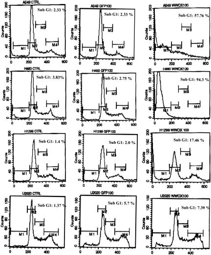
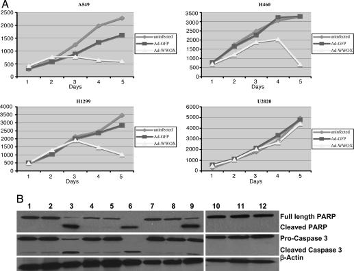
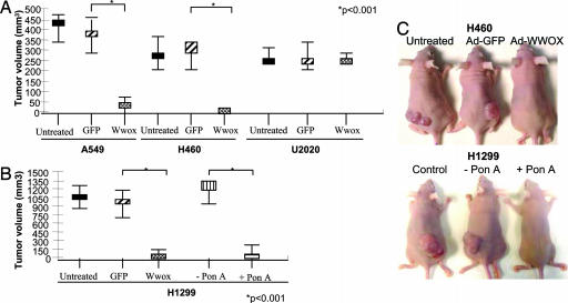
The WWOX (WW domain containing oxidoreductase) gene at the common fragile site, FRA16D, is altered in many types of cancer, including lung cancer. We have examined the tumor suppressor function of WWOX in preclinical lung cancer models. The WWOX gene was expressed in lung cancer cell lines through recombinant adenovirus (Ad) infection (Ad-WWOX), and through a drug [ponasterone A, (ponA)]-inducible system. After WWOX restoration in vitro, endogenous Wwox protein-negative cell lines (A549, H460, and H1299) underwent apoptosis through activation of the intrinsic apoptotic caspase cascade in A549 and H460 cells. Ectopic expression of Wwox caused dramatic suppression of tumorigenicity of A549, H460, and H1299 cells in nude mice after Ad-WWOX infection and after ponA induction of Wwox expression in H1299 lung cancer cells. Tumorigenicity and in vitro growth of U2020 (Wwox-positive) lung cancer cells was unaffected by Wwox overexpression. This study confirms that WWOX is a tumor suppressor gene and is highly effective in preventing growth of lung cancer xenografts, whether introduced through viral infection or by induction of a silent WWOX transgene.


Figures

Fig. 1.
Fig. 1.
Expression of Wwox protein. (A) Expression of endogenous Wwox is detected in U2020 and MCF7 cells but not in H1299, H460, or A549 cells (50 μg of proteins loaded). Lane 1, H1299; lane 2, H460; lane 3, A549; lane 4, U2020; lane 5, MCF-7. (B) Expression of Wwox after infection with Ad-WWOX (25 μg loaded). Lane 1, H1299, Ad-WWOX-infected; lane 2, H1299, Ad-GFP-infected; lane 3, H1299; lane 4, H460, Ad-WWOX-infected; lane 5, H460, Ad-GFP-infected; lane 6, H460; lane 7, A549, Ad-WWOX-infected; lane 8, A549, Ad-GFP-infected; lane 9, A549.
Fig. 2.
Fig. 2.
Flow cytometry analysis of untreated, Ad-GFP-, and Ad-WWOX-infected cells. Wwox-negative A549, H460, and H1299 cells undergo apoptosis 5 days after restoration of Wwox expression by Ad-WWOX infection, but U2020 cells are unaffected. Ad-GFP infection did not induce apoptosis.
Fig. 3.
Fig. 3.
Effect of Wwox expression on cell growth in vitro. (A) Growth of uninfected, Wwox-negative A549, H460, and H1299 cells, and cells after infection with Ad-GFP and Ad-WWOX. (B) Immunoblot detection of PARP and caspase 3. Lane 1, A549; lane 2, A549/Ad-GFP; lane 3, A549/Ad-WWOX; lane 4, H460; lane 5, H460/Ad-GFP; lane 6, H460/Ad-Wwox; lane 7, H1299; lane 8, H1299/Ad-GFP; lane 9, H1299/Ad-WWOX; lane 10, U2020; lane 11, U2020/Ad-GFP; lane 12, U2020/Ad-WWOX. PARP is cleaved in Wwox-negative cell lines when Wwox is restored through Ad-Wwox infection (lanes 3, 6, and 9). Caspase 3 is cleaved in A549 and H460 (lanes 3 and 6) but not in H1299 cells after Ad-WWOX infection. In U2020 cells, neither PARP nor caspase 3 is cleaved after Ad-WWOX infection (lane 12).
Fig. 4.
Fig. 4.
Inducible expression of Wwox in H1299/I cells. (A) Cells were cultured in the presence (+) or absence (–) of 10 μM ponA for 48 hr and tested for Wwox expression. Clones 7 and 2, which expressed the transgene only upon induction with ponA, were used in subsequent experiments. GAPDH expression servedas loading control. (B) H1299/I clone 7 cells incubated in the absence or presence of increasing concentrations of ponA for 48 hr. Wwox levels increased in a dose-dependent manner and were quantified by densitometry, normalized to GAPDH expression levels. (C) Time course of Wwox induction in H1299/I clone 7 cells after treatment with 10 μM ponA. Wwox levels were quantified by densitometry. (D) Effect of 10 μM ponA on growth of H1299/I clone 7 cells. On day 1, ponA was added, and maximum Wwox expression was found on day 4. From day 5, the induced cells (H1299/I+) grow significantly more slowly than uninduced cells (H1299/I)(P < 0.001). The experiment was done in triplicate.
Fig. 5.
Fig. 5.
Effect of Wwox expression on tumorigenicity of lung cancer cells. (A) Tumor volume of untreated, Ad-GFP-, and Ad-WWOX-infected A549, H460, and U2020 lung cancer cells. Restoration of Wwox expression in A549 and H460 cells suppressed tumor growth significantly (P < 0.001) compared with Ad-GFP infected cells. (B) Tumor volume of untreated, Ad-GFP-, and Ad-WWOX-infected H1299 cells and H1299/I and H1299/I+ cells. Tumors were suppressed in Ad-WWOX-infected H1299 cells and in H1299/I+ cells. (C) Examples of tumor formation by uninfected, Ad-GFP-, and Ad-WWOX-infected A549, H1299/I, and H1299/I+ cells.
Fig. 6.
Fig. 6.
Ex vivo analysis of H1299/I and H1299/I+ cells. (A) Protein lysates from H1299 (lane 1), uninduced H1299/I (lanes 2, 3, and 4), and induced H1299/I+ (lane 5) tumors tested for Wwox expression by immunoblot analysis. Wwox was not expressed in the H1299/I or H1299/I+ tumors. (B) A portion of the H1299I/+ tumor was plated and cultured, and cells were treated with ponA. Wwox was reexpressed after 48 hr of treatment with 10 μM ponA, indicating the presence of the inducible WWOX plasmid.

Similar articles

Cited by

References

    1. Greenlee, R. T., Hill-Harmon, M. B., Murray, T. & Thun, M. (2001) CA Cancer J. Clin. 51, 15–36. - PubMed
    1. Roth, J. A., Nguyen, D., Lawrence, D. D., Kemp, B. L., Carrasco, C. H., Ferson, D. Z., Hong, W. K., Komaki, R., Lee, J. J., Nesbitt, J. C., et al. (1996) Nat. Med. 2, 985–991. - PubMed
    1. Nemunaitis, J., Swisher, S. G., Timmons, T., Connors, D., Mack, M., Doerksen, L., Weill, D., Wait, J., Lawrence, D. D., Kemp, B. L., et al. (2000) J. Clin. Oncol. 18, 609–622. - PubMed
    1. Roth, J. A., Swisher, S. G., Merritt, J. A., Lawrence, D. D., Kemp, B. L., Carrasco, C. H., El-Naggar, A. K., Fossella, F. V., Glisson, B. S., Hong, W. K., et al. (1998) Semin. Oncol. 25, Suppl. 8, 33–37. - PubMed
    1. Weill, D., Mack, M., Roth, J., Swisher, S., Proksch, S., Merritt, J. & Nemunaitis, J. (2000) Chest 118, 966–970. - PubMed

Publication types