. 2005 Oct 13;33(18):e160.
doi: 10.1093/nar/gni158.

A simple method for displaying recalcitrant proteins on the surface of bacteriophage lambda

Affiliations

A simple method for displaying recalcitrant proteins on the surface of bacteriophage lambda

Christine N Zanghi et al. Nucleic Acids Res. .

Abstract

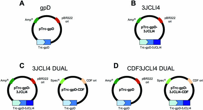
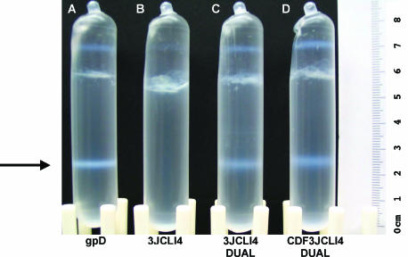
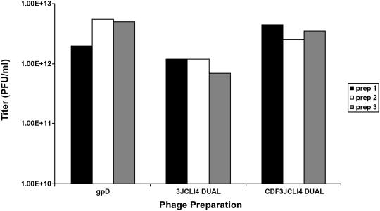
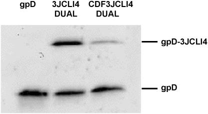
Bacteriophage lambda (lambda) permits the display of many foreign peptides and proteins on the gpD major coat protein. However, some recombinant derivatives of gpD are incompatible with the assembly of stable phage particles. This presents a limitation to current lambda display systems. Here we describe a novel, plasmid-based expression system in which gpD deficient lambda lysogens can be co-complemented with both wild-type and recombinant forms of gpD. This dual expression system permits the generation of mosaic phage particles that contain otherwise recalcitrant recombinant gpD fusion proteins. Overall, this improved gpD display system is expected to permit the expression of a wide variety of peptides and proteins on the surface of bacteriophage lambda and to facilitate the use of modified lambda phage vectors in mammalian gene transfer applications.


Figures

Figure 1
Figure 1
gpD Expression constructs. Plasmid vectors for prokaryotic expression of wild-type and recombinant forms of gpD are shown. (A) An ECO derivative of gpD (blue) was inserted into an ampicillin-selectable, pBR322-based pTrc expression plasmid, where it was placed under the transcriptional control of the high-level, regulatable Trc promoter (light blue). (B) A sequence coding for a high-affinity αvβ3 binding protein derived from the tenth fibronectin type III domain (3JCLI4; dark blue) was inserted at the 3′ end of the gpD insert in the plasmid shown in (A), resulting in the generation of a construct that encoded a gpD-3JCLI4 fusion protein. (C) A matched dual expression plasmid set, capable of co-expressing wild-type gpD and the gpD-3JCLI4 fusion protein is shown. The ampicillin selection marker (dark green) and pBR322-derived (dark red) origin of replication in the plasmid shown in (A) were replaced with a spectinomycin resistance gene (light green) and a pCDF1-derived origin of replication (light red), to generate the gpD-encoding plasmid shown on the left. (D) A second matched dual expression plasmid set, capable of co-expressing wild-type gpD and the gpD-3JCLI4 fusion protein is shown. In this case, the ampicillin selection marker and pBR322-derived origin of replication in the plasmid shown in (B) were replaced with a spectinomycin resistance gene and a pCDF1-derived origin of replication, to generate the gpD-3JCLI4-encoding plasmid shown on the right.
Figure 2
Figure 2
Co-expression of 3JCLI4-gpD and wild-type gpD yields intact phage particles. Lysogens of TOP10 cells containing gpD-deficient λ D1180 (Luc) were transformed with single plasmid vectors encoding either wild-type gpD (gpD; see Figure 1A) or the recombinant gpD-3JCLI4 fusion protein (3JCLI4; see Figure 1B). Alternatively, the cells were co-transformed with two plasmids, corresponding to the constructs shown in Figure 1C (3JCLI4 DUAL) or Figure 1D (CDF3JCLI4 DUAL); these paired constructs permitted the co-expression of wild-type and recombinant gpD in the same E.coli host cell. Following lysogen induction and cell lysis, phage particles were pelleted and subjected to cesium chloride density gradient purification. The results are shown; the large arrow denotes the characteristic λ phage band (the upper bands correspond largely to non-infectious protein debris; data not shown). It can be readily appreciated that stable phage particles were recovered from all of the preparations, except for the phage that were exclusively complemented by the recombinant gpD-3JCLI4 protein. This suggests that the gpD-3JCLI4 protein failed to support the assembly of stable phage particles and that this deficiency could be overcome by co-expression of the wild-type gpD protein (as in the 3JCLI4 DUAL and CDF3JCLI4 DUAL preparations).
Figure 3
Figure 3
Co-expression of 3JCLI4-gpD and wild-type gpD yields infectious phage particles. Lysogens of TOP10 cells containing gpD-deficient λ D1180 (Luc) were transformed with plasmid vectors encoding either wild-type gpD alone (gpD; see Figure 1A) or with two plasmids, corresponding to the constructs shown in Figure 1C (3JCLI4 DUAL) or Figure 1D (CDF3JCLI4 DUAL); these paired constructs permitted the co-expression of wild-type and recombinant gpD in the same E.coli host cell. Following lysogen induction, CsCl-banding and dialysis, purified phage particles were titered on LE392 E.coli host cells. Results shown represent phage titers from three separate phage preparations; no appreciable amounts of infectious phage were recovered from lysogens that expressed only the gpD-3JCLI4 fusion protein (Figure 1B).
Figure 4
Figure 4
Co-expression of 3JCLI4-gpD and wild-type gpD results in the generation of mosaic phage particles. Lysogens of TOP10 cells containing gpD-deficient λ D1180 (Luc) were transformed with plasmid vectors encoding either wild-type gpD alone (gpD) or with two plasmids that permitted the co-expression of wild-type and recombinant gpD in the same E.coli host cell (3JCLI4 DUAL and CDF3JCLI4 DUAL; see Figures 1–3). Following lysogen induction, CsCl-banding and dialysis, purified phage particles were titered on LE392 E.coli host cells. A total of 1 × 10⁹ PFU of each preparation was then loaded on a 20% SDS–PAGE gel and phage protein content was examined by immunoblot analysis, using a rabbit polyclonal antiserum directed against gpD. The results show incorporation of wild-type gpD (∼12 kDa) into all of the phage preparations (as expected), as well as inclusion of recombinant gpD-3JCLI4 (∼23 kDa) in those preparations that were generated from lysogens that had been co-transformed with both wild-type and recombinant gpD expression vectors. It is also apparent that lower levels of recombinant gpD-3JCLI4 were incorporated when the recombinant protein was expressed from a CDF-origin containing construct, as compared to a pBR322-based plasmid.

Similar articles

Cited by

References

    1. Murialdo H., Ray P.N. Model for arrangement of minor structural proteins in head of bacteriophage lambda. Nature. 1975;257:815–817. - PubMed
    1. Sternberg N., Weisberg R. Packaging of coliphage lambda DNA. II. The role of the gene D protein. J. Mol. Biol. 1977;117:733–759. - PubMed
    1. Yang F., Forrer P., Dauter Z., Conway J.F., Cheng N., Cerritelli M.E., Steven A.C., Pluckthun A., Wlodawer A. Novel fold and capsid-binding properties of the lambda-phage display platform protein gpD. Nat. Struct. Biol. 2000;7:230–237. - PubMed
    1. Sternberg N., Hoess R.H. Display of peptides and proteins on the surface of bacteriophage lambda. Proc. Natl Acad. Sci. USA. 1995;92:1609–1613. - PMC - PubMed
    1. Smith M.P., Feiss M. Sites and gene products involved in lambdoid phage DNA packaging. J. Bacteriol. 1993;175:2393–2399. - PMC - PubMed

Publication types