. 2005 Oct 13;33(18):e161.
doi: 10.1093/nar/gni162.

Method for multiplex cellular detection of mRNAs using quantum dot fluorescent in situ hybridization

Affiliations

Method for multiplex cellular detection of mRNAs using quantum dot fluorescent in situ hybridization

PokMan Chan et al. Nucleic Acids Res. .

Abstract

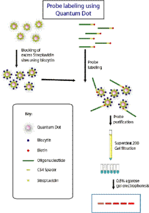
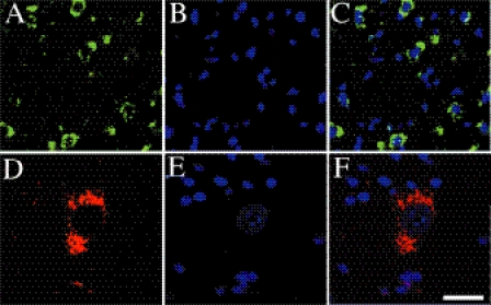
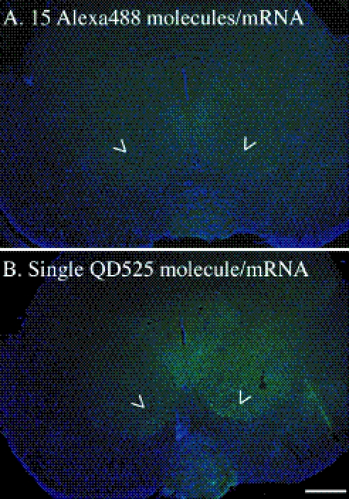
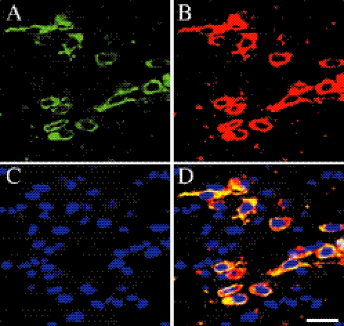
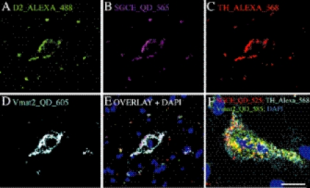
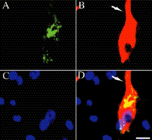
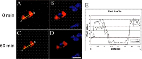
The photostability and narrow emission spectra of non-organic quantum dot fluorophores (QDs) make them desirable candidates for fluorescent in situ hybridization (FISH) to study the expression of specific mRNA transcripts. We developed a novel method for direct QD labeling of modified oligonucleotide probes through streptavidin and biotin interactions, as well as protocols for their use in multiple-label FISH. We validated this technique in mouse brainstem sections. The subcellular localization of the vesicular monoamine transporter (Vmat2) mRNA corresponds when using probes labeled with two different QDs in the same hybridization. We developed protocols for combined direct QD FISH and QD immunohistochemical labeling within the same neurons as well as for simultaneous study of the subcellular distribution of multiple mRNA targets. We demonstrated increased sensitivity of FISH using QDs in comparison with organic fluorophores. These techniques gave excellent histological results both for multiplex FISH and combined FISH and immunohistochemistry. This approach can facilitate the ultrasensitive simultaneous study of multiple mRNA and protein markers in tissue culture and histological section.


Figures

Figure 1
Figure 1
(A) Schematic representation of quantum dot oligonucleotide probe labeling strategy for FISH. Excess streptavidin sites on the QDs were first blocked by biocytin before adding to the biotinylated oligonucleotide for labeling. The QD labeled oligonucleotide probes were purified using a Superdex 200 column.
Figure 2
Figure 2
Agarose gel electrophoretic analysis of biotinylated oliogonucleotide probes with streptavidin-conjugated QDs 585. The individual samples represent fractions eluted from a Superdex 200 column. The Superdex 200 purified oligonucleotide was used directly for FISH. High-molecular weight conjugates eluted in later fractions (data not shown).
Figure 3
Figure 3
Fluorescent labeling of cellular mRNA in mouse brain sections using QDs. (A) Dopaminergic cells in SN labeled with Vmat2 oligonucleotide containing QD 525. (B) DAPI staining of nuclei. (C) Overlay of the QD 525 and DAPI signals. (D) High magnification image of cell in SN labeled with Vmat2 QD 585 oligonucleotide probe. Note the specific cytoplasmic labeling of the Vmat2 mRNA and the low tissue background. The images were obtained from 25 µm tissue sections and captured using a ×100/1.3 NA objective with ×4 digital zoom. Simultaneous detection of DAPI, QD 525 and QD 585 signals were excited by a diode 405 laser on a Zeiss Meta LSM. Scale bar = 25 µm for (A–C) and 10 µm for (DF).
Figure 4
Figure 4
Comparison of organic fluorophore and QD FISH sensitivity. (A) Mouse midbrain section hybridized with three probes for ɛ-sarcoglycan mRNA (SGCE) each labeled with five Alexa 488 fluorophores. In our hands, this protocol gives excellent histological results for ×20 images of this mouse brain region, as described previously (8). However, at ×4 magnification, neurons that are known to express this mRNA within the mouse red nucleus are not able to be detected (arrow heads). Alexa signal is green and DAPI nuclear counterstain is blue. (B) The same region studied with one of the three ɛ-sarcoglycan mRNA probe sequences used in (A), labeled with a single QD 525 fluorophore. Neurons in the red nucleus that express this mRNA are clearly visible (arrow heads). QD 525 signal is green and DAPI signal is blue. Scale bar = 500 µm.
Figure 5
Figure 5
Double labeling FISH using QD 525 and QD 585 oligonucleotide probes. Neurons in SN were labeled with two different QD oligonucleotide probes. Vmat2 mRNA was detected using both QD 525 (A) and QD 585 (B) probes. The fluorescent signals for DAPI (C) and QDs were detected simultaneously using a diode 405 blue laser of the Zeiss Meta 510 LSM. The FISH signal was detected only in cytoplasm and low background signal. Note the complete overlap of QD 525 and QD 585 signal (D). Scale bar = 20 µm.
Figure 6
Figure 6
Multiple-label and multi-spectral FISH using QD and organic fluorophores. Probes were labeled with the fluorophores indicated and hybridization for multiple mRNA targets was performed. (AD) Detection of four different mRNA targets separated by spectral imaging of a 0.2 µm optical section. The emission of each individual fluorophore was clearly separated. (E) Overlay of (A–D). (F) Overlay of higher magnification spectral imaging of a single neuron labeled for three different mRNAs. Note the differences in the subcellular pattern of each mRNA target. DAPI counterstaining of the same sections is blue. Scale bar: (A–E) = 20 µm; (F) = 10 µm.
Figure 7
Figure 7
Simultaneous multiplex labeling of mRNA and protein in mouse brain using QD 525 and QD 605. In situ hybridization was performed and the same tissue sections were processed for immunohistochemistry using QD for the detection of protein epitopes. (A) Vmat2 mRNA positive neurons in SN was labeled with QD 525. (B) The same cell was labeled with TH antibody and the signal was detected using streptadvidin conjugated QD 605. Note the labeling of both Vmat2 mRNA and TH protein in the same cell. The Vmat2 mRNA signal is restricted to the cytoplasm whereas the labeling of TH is extended to the whole cell body and processes (arrow). (C) DAPI labeling of nuclei. (D) Overlay shows the differing subcellular distributions of Vmat2 mRNA and TH immunoreactivity. Scale bar = 15 µm.
Figure 8
Figure 8
QD FISH probe is resistant to photobleaching. (A, B) A neuron in DMX was labeled with QD 585 and the image was captured using the 405 nm laser at time 0 min. (C,D) The cell was continuously irradiated by the blue laser for another 60 min and the image was captured. The settings for laser power, gain, offset, scanning time and pinhole were identical for both images and the fluorescence emission was recorded through a ×100 NA 1.3 oil objective. (E) Fluorescence intensity across the same cell coordinate was measured using NIH Image J program. Scale bar: (A–D) = 15 µm.

References

    1. Burgunder J.M., Young W.S., III The distribution of thalamic projection neurons containing cholecystokinin messenger RNA, using in situ hybridization histochemistry and retrograde labeling. Brain Res. 1988;464:179–189. - PubMed
    1. Forghani B., Dupuis K.W., Schmidt N.J. Rapid detection of herpes simplex virus DNA in human brain tissue by in situ hybridization. J. Clin. Microbiol. 1985;22:656–658. - PMC - PubMed
    1. Durrant I., Brunning S., Eccleston L., Chadwick P., Cunningham M. Fluorescein as a label for non-radioactive in situ hybridization. Histochem. J. 1995;27:94–99. - PubMed
    1. Hermanson O., Ericson H., Sanchez-Watts G., Watts A.G., Blomqvist A. Autoradiographic visualization of 35S-labeled cRNA probes combined with immunoperoxidase detection of choleragenoid: a double-labeling light microscopic method for in situ hybridization and retrograde tract tracing. J. Histochem. Cytochem. 1994;42:827–831. - PubMed
    1. Chartrand P., Bertrand E., Singer R.H., Long R.M. Sensitive and high-resolution detection of RNA in situ. Methods Enzymol. 2000;318:493–506. - PubMed

Publication types

MeSH terms

Substances