

Immunoexpression of HBME-1, high molecular weight cytokeratin, cytokeratin 19, thyroid transcription factor-1, and E-cadherin in thyroid carcinomas
- PMID: 16224162
- PMCID: PMC2779285
- DOI: 10.3346/jkms.2005.20.5.853
Immunoexpression of HBME-1, high molecular weight cytokeratin, cytokeratin 19, thyroid transcription factor-1, and E-cadherin in thyroid carcinomas
Abstract
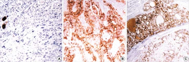
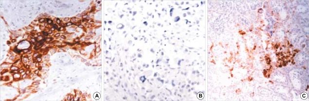
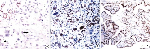
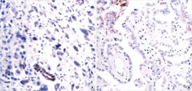
To examine the immunohistochemical alterations associated with the histological dedifferentiation of thyroid carcinomas, we performed staining for HBME-1, high molecular weight cytokeratin (HCK), CK 19, thyroid transcription factor-1 (TTF-1) and E-cadherin (E-CD) on 125 various types of thyroid carcinomas. The HBME-1 staining was strong and diffuse in follicular carcinoma (FC), papillary carcinoma (PC), and poorly differentiated carcinoma (PDC), while it was rare in undifferentiated carcinoma (UC) as well as in benign lesions. Strong, diffuse staining for CK19 and HCK was predominantly found in PC, and these markers were not much found in other carcinomas. TTF-1 uniformly stained the tumor cells of all cases of PC, FC and Hurthle cell carcinoma (HC) and 42% of the PDC, while there was only focal staining in one case of the UC. Compared to the strong, diffuse reactivity in the benign lesions, E-CD staining was noted in 67% of PC, 80% of FC, 83% of HC, 58% of PDC and none of the UC. These results suggest that HBME-1 may be a marker for well-differentiated carcinomas while CK19 and HCK are phenotypic markers for papillary carcinoma. The loss or reduced expression of TTF-1 and E-CD may be markers for dedifferentiation.
Figures
Similar articles
-
Immunohistochemical diagnosis of papillary thyroid carcinoma.Mod Pathol. 2001 Apr;14(4):338-42. doi: 10.1038/modpathol.3880312. Mod Pathol. 2001. PMID: 11301350
-
Diagnostic value of galectin-3, HBME-1, cytokeratin 19, high molecular weight cytokeratin, cyclin D1 and p27(kip1) in the differential diagnosis of thyroid nodules.J Korean Med Sci. 2007 Aug;22(4):621-8. doi: 10.3346/jkms.2007.22.4.621. J Korean Med Sci. 2007. PMID: 17728499 Free PMC article.
-
Comparison of proliferating cell nuclear antigen, thyroid transcription factor-1, Ki-67, p63, p53 and high-molecular weight cytokeratin expressions in papillary thyroid carcinoma, follicular carcinoma, and follicular adenoma.Ann Diagn Pathol. 2011 Apr;15(2):108-16. doi: 10.1016/j.anndiagpath.2010.11.005. Ann Diagn Pathol. 2011. PMID: 21315633
-
Value of thyroid transcription factor-1 immunostaining in tumor diagnosis: a review and update.Appl Immunohistochem Mol Morphol. 2012 Oct;20(5):429-44. doi: 10.1097/PAI.0b013e31825439bc. Appl Immunohistochem Mol Morphol. 2012. PMID: 22531688 Review.
-
Thyroid transcription factor-1: a review.Appl Immunohistochem Mol Morphol. 2002 Jun;10(2):97-102. doi: 10.1097/00129039-200206000-00001. Appl Immunohistochem Mol Morphol. 2002. PMID: 12051643 Review.
Cited by
-
Universal markers of thyroid malignancies: galectin-3, HBME-1, and cytokeratin-19.Endocr Pathol. 2010 Jun;21(2):80-9. doi: 10.1007/s12022-010-9114-y. Endocr Pathol. 2010. PMID: 20198455
-
The Combined Utility of HBME-1 and Galectin-3 Immunohistochemistry and BRAF V600E Mutations in the Diagnosis of Papillary Thyroid Carcinoma.Cureus. 2021 Dec 10;13(12):e20339. doi: 10.7759/cureus.20339. eCollection 2021 Dec. Cureus. 2021. PMID: 34934597 Free PMC article.
-
CK19 IS A USEFUL MARKER IN DISTINGUISHING FOLLICULAR VARIANT OF PAPILLARY THYROID CARCINOMA FROM BENIGN THYROID LESIONS WITH FOLLICULAR GROWTH PATTERN.Acta Endocrinol (Buchar). 2016 Oct-Dec;12(4):387-391. doi: 10.4183/aeb.2016.387. Acta Endocrinol (Buchar). 2016. PMID: 31149120 Free PMC article.
-
Diagnostic utility of immunohistochemical panel in various thyroid pathologies.Langenbecks Arch Surg. 2010 Sep;395(7):885-91. doi: 10.1007/s00423-010-0690-6. Epub 2010 Jul 18. Langenbecks Arch Surg. 2010. PMID: 20640858
-
E-cadherin expression and cell proliferation in the primary tumor and metastatic lymph nodes of papillary thyroid microcarcinoma.Mol Clin Oncol. 2014 Mar;2(2):226-232. doi: 10.3892/mco.2013.220. Epub 2013 Dec 5. Mol Clin Oncol. 2014. PMID: 24649337 Free PMC article.
References
-
- Rosai J. Thyroid gland. In: Rosai J, editor. Rosai and Ackermann's Surgical Pathology. 9 ed. Phildelphia: Elsevier; 2004. pp. 515–594.
-
- van Hoeven KH, Kovatich AJ, Miettinen M. Immunocytochemical evaluation of HBME-1, CA 19-9, and CD-15 (Leu-M1) in fine-needle aspirates of thyroid nodules. Diagn Cytopathol. 1998;18:93–97. - PubMed
-
- Casey MB, Lohse CM, Lloyd RV. Distinction between papillary thyroid hyperplasia and papillary thyroid carcinoma by immunohistochemical staining for cytokeratin 19, galectin-3, and HBME-1. Endocr Pathol. 2003;14:55–60. - PubMed
-
- Sack MJ, Astengo-Osuna C, Lin BT, Battifora H, LiVolsi VA. HBME-1 immunostaining in thyroid fine-needle aspirations: a useful marker in the diagnosis of carcinoma. Mod Pathol. 1997;10:668–674. - PubMed
-
- Cameron BR, Berean KW. Cytokeratin subtypes in thyroid tumors: immunohistochemical study with emphasis on the follicular variant of papillary carcinoma. J Otolaryngol. 2003;32:319–322. - PubMed
Publication types
MeSH terms
Substances
LinkOut - more resources
Full Text Sources
Medical
Research Materials
