Clinical Trial
. 2005 Nov-Dec;28(6):582-92.
doi: 10.1097/01.cji.0000175468.19742.10.

Inability of a fusion protein of IL-2 and diphtheria toxin (Denileukin Diftitox, DAB389IL-2, ONTAK) to eliminate regulatory T lymphocytes in patients with melanoma

Affiliations
Clinical Trial

Inability of a fusion protein of IL-2 and diphtheria toxin (Denileukin Diftitox, DAB389IL-2, ONTAK) to eliminate regulatory T lymphocytes in patients with melanoma

Peter Attia et al. J Immunother. 2005 Nov-Dec.

Abstract

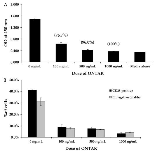
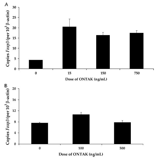
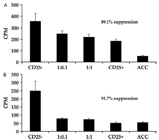
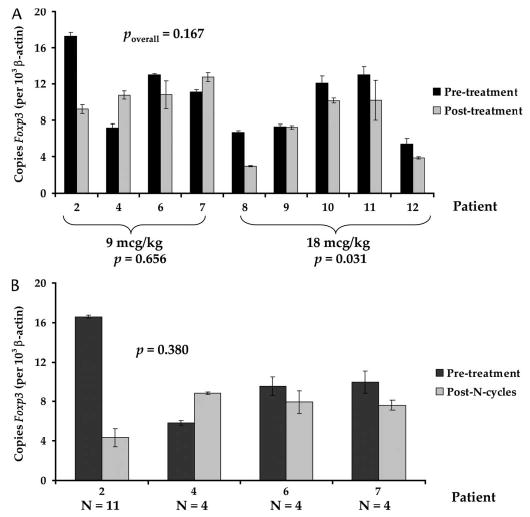
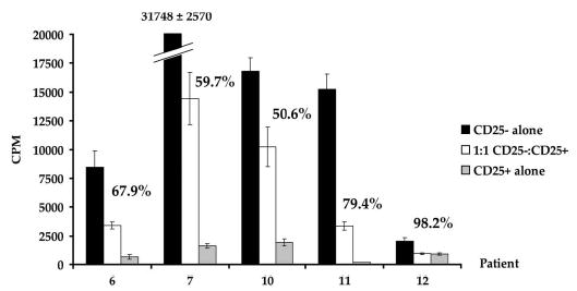
Elimination of regulatory T lymphocytes may provide a way to break self-tolerance and unleash the anti-tumor properties of circulating lymphocytes. The use of fusion proteins, which link cytotoxic molecules to receptor targets, provides one approach to this problem. This study examined the ability of a fusion protein of interleukin-2 (IL-2) and diphtheria toxin (Denileukin Diftitox, DAB389IL-2, ONTAK) to eliminate regulatory T lymphocytes based on their expression of high-affinity IL-2 receptors. Thirteen patients (12 with metastatic melanoma, 1 with metastatic renal cell carcinoma) were treated at one of the two Food and Drug Administration-approved doses of Denileukin Diftitox (seven patients at 9 microg/kg, six patients at 18 microg/kg). None of the patients experienced an objective clinical response. Foxp3 expression did not decrease significantly overall, although it did decrease minimally among patients receiving 18 microg/kg (-2.01+/-0.618 copies of Foxp3/10(3) copies of beta-actin; P=0.031). Denileukin Diftitox did not decrease the suppressive ability of CD4CD25 cells as quantified by an in vitro co-culture suppression assay. Furthermore, the increased numbers of lymphocytes in patients resulting from treatment with IL-2 were not susceptible to Denileukin Diftitox. Administration of Denileukin Diftitox does not appear to eliminate regulatory T lymphocytes or cause regression of metastatic melanoma.


Figures

FIGURE 1
FIGURE 1
A, WST-1 assay on Atac4 cell line that expressed high levels of CD25 following treatment with Denileukin Diftitox in vitro for 72 hours. Numbers in parentheses above columns indicate the percentage decrease in optical density relative to untreated group. Uptake of a blank well (medium alone) is shown for comparison. In vitro, Denileukin Diftitox eliminated cells expressing CD25. B, PBMCs treated with 50 ng/mL anti-CD3 for 72 hours, prior to 72-hour in vitro exposure to Denileukin Diftitox. Viability was assessed by failure to take up PI stain. The percentage of cells expressing CD25 decreased with increasing doses of Denileukin Diftitox, and this was paralleled by cell viability, demonstrating that Denileukin Diftitox could eliminate activated PBMCs in vitro.
FIGURE 2
FIGURE 2
A, Foxp3 expression in purified CD4+ cells following 48-hour in vitro exposure to Denileukin Diftitox did not demonstrate evidence of Denileukin Diftitox–mediated elimination of Foxp3 expressing regulatory T cells. B, Foxp3 expression in purified CD4+ cells following 72-hour in vitro exposure to Denileukin Diftitox also failed to demonstrate elimination of Foxp3 expressing regulatory T cells.
FIGURE 3
FIGURE 3
Suppressive capacity of untreated CD4+CD25+ purified T cells (A) and Denileukin Diftitox–treated CD4+CD25+ purified T cells (500 ng/mL × 72 hours) (B). Percent suppression reflects suppression for 1:1 treatment group (10,000 CD25:10,000 CD25+ cells). Treatment with Denileukin Diftitox did not reduce suppressive abilities of CD4+CD25+ in vitro. ACC, T cell–depleted irradiated accessory cell.
FIGURE 4
FIGURE 4
A, Peripheral blood counts from four patients during the first two to three treatment cycles. Arrows along x axis denote the first day of treatment of a given 5-day cycle. Treatment with Denileukin Diftitox caused an abrupt fall in ALC and rise in ANC, both of which were transient changes. B, Peripheral blood counts for patient 2 over the entire course of his 11 cycles. Arrows along x axis denote the first day of treatment of a given 5-day cycle. With successive treatments, the magnitude of change of both the ALC and the total WBC decreased.
FIGURE 5
FIGURE 5
A, Change in Foxp3 expression in purified CD4+ cells following in vivo administration of Denileukin Diftitox after the first cycle of treatment of nine patients. The four patients (2, 4, 6, 7) who received 9 μg/kg did not demonstrate a significant change in Foxp3 expression, whereas the five patients (8, 9, 10, 11, 12) who received 18 μg/kg experienced a small but statistically significant decrease in Foxp3 expression, suggesting that Denileukin Diftitox had minimal impact on Foxp3 expression in vivo. P values reflect two-tailed paired t test. B, Change in Foxp3 expression in purified CD4+ cells following multiple cycles of Denileukin Diftitox. Foxp3 expression did not change significantly following multiple treatments. N = number of cycles. P value reflects two-tailed paired t test.
FIGURE 6
FIGURE 6
Suppressive ability of CD4+CD25+ freshly isolated T cells from five patients following two cycles of Denileukin Diftitox. In each patient, at least 50% suppressive activity was measured following treatment, suggesting that Denileukin Diftitox failed to eliminate T cells capable of mediating suppression.
FIGURE 7
FIGURE 7
Foxp3 expression in purified CD4+ cells from two patients treated with high-dose IL-2. Cells were harvested 3 days following last dose of IL-2, during rebound lymphocytosis, and treated with 0, 100, or 1000 ng/mL Denileukin Diftitox in vitro. These IL-2-activated regulatory T cells were not more sensitive to Denileukin Diftitox treatment in vitro.

References

    1. Kawaida H, Kono K, Takahashi A, et al. Distribution of CD4+CD25 high regulatory T-cells in tumor-draining lymph nodes in patients with gastric cancer. J Surg Res. 2005;124:151–157. - PubMed
    1. Lundgren A, Stromberg E, Sjoling A, et al. Mucosal Foxp3-expressing CD4+ CD25 high regulatory T cells in Helicobacter pylori-infected patients. Infect Immun. 2005;73:523–531. - PMC - PubMed
    1. Jonuleit H, Schmitt E. Regulatory T-cells in antitumor therapy: Isolation and functional testing of CD4+CD25+ regulatory T-cells. Methods Mol Med. 2005;109:285–296. - PubMed
    1. Karube K, Ohshima K, Tsuchiya T, et al. Expression of Foxp3, a key molecule in CD4CD25 regulatory T cells, in adult T-cell leukaemia/lymphoma cells. Br J Haematol. 2004;126:81–84. - PubMed
    1. Wang HY, Lee DA, Peng G, et al. Tumor-specific human CD4+ regulatory T cells and their ligands: Implications for immunotherapy. Immunity. 2004;20:107–118. - PubMed

Publication types

MeSH terms