. 2005 Nov;115(11):3239-49.
doi: 10.1172/JCI24731. Epub 2005 Oct 13.

Human skin cells support thymus-independent T cell development

Affiliations

Human skin cells support thymus-independent T cell development

Rachael A Clark et al. J Clin Invest. 2005 Nov.

Abstract

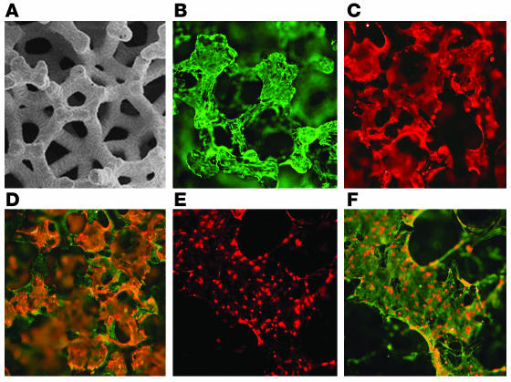
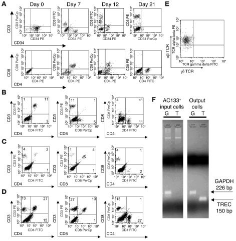
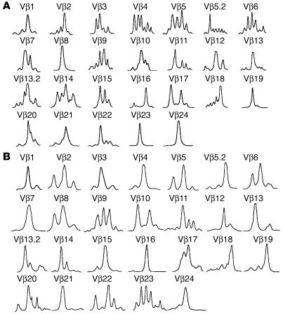
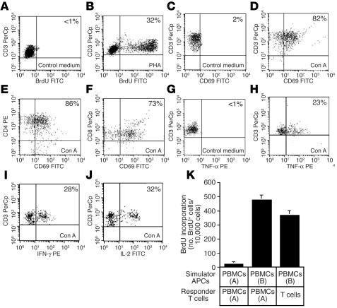
Thymic tissue has previously been considered a requirement for the generation of a functional and diverse population of human T cells. We report that fibroblasts and keratinocytes from human skin arrayed on a synthetic 3-dimensional matrix support the development of functional human T cells from hematopoietic precursor cells in the absence of thymic tissue. Newly generated T cells contained T cell receptor excision circles, possessed a diverse T cell repertoire, and were functionally mature and tolerant to self MHC, indicating successful completion of positive and negative selection. Skin cell cultures expressed the AIRE, Foxn1, and Hoxa3 transcription factors and a panel of autoantigens. Skin and bone marrow biopsies can thus be used to generate de novo functional and diverse T cell populations for potential therapeutic use in immunosuppressed patients.


Figures

Figure 1
Figure 1
Structure of 3-dimensional skin cell cultures. (A) Scanning electron micrograph of the Cellfoam 3-dimensional matrix. Image courtesy of Cytomatrix LLC. (B and C) IF demonstrating the morphologies of (B) fibroblasts stained with vimentin antibody and (C) keratinocytes stained with antibodies to cytokeratins when these cells were grown alone on matrices. (D) When grown together, keratinocytes (orange) and fibroblasts (green) occupied distinct sites on the matrices. (E) DCs, identified by intense staining with HLA-DR antibodies, were observed only if bone marrow progenitor cells were added to the matrices. (F) DCs were often found adherent to the surface of vimentin+ fibroblasts. Magnification: ×2 (E and F), ×5 (A), ×10 (BD).
Figure 2
Figure 2
HPCs differentiate into T cells in skin cell cultures. (A) Maturation of surface markers during T cell development in skin cell cultures. Matrices were irradiated prior to the addition of HPCs and produced primarily CD4+ T cells. (BD) Alterations in culture conditions influenced the production of CD4+ versus CD8+ cells. (B) Irradiation of the construct prior to the addition of HPCs supported differentiation of proportionately more CD4+ cells. (C) Treatment of the skin cell construct with IFN-γ before the addition of HPCs increased the percentage of CD8+ cells produced. (D) In the absence of these treatments, a more equal distribution of CD4+ and CD8+ cells was observed. Cells were harvested from the matrices at 28 days. Similar results were seen in duplicate experiments. The percentage of cells in each quadrant is shown. (E) Expression of αβ TCR versus γδ TCR by CD3+ cells produced in skin cell cultures. (F) Qualitative RT-PCR for expression of TREC (T) and GAPDH (G) of input AC133+ HPCs and output T cells produced in skin cell cultures.
Figure 3
Figure 3
Spectratype analysis of T cells produced in skin cell cultures. T cells generated in skin cell cultures were subjected to TCR-CDR3 length analysis. Diversity within each Vβ family is signified by multiple peaks. Precursor cells from 1 bone marrow donor were matured in skin cell cultures from 2 different skin donors. T cells produced in cultures from the first skin donor (A; 8 × 10⁵ cells analyzed) were more diverse and had a different T cell repertoire than T cells produced in cultures from the second skin donor (B; 5 × 10⁵ cells analyzed).
Figure 4
Figure 4
T cells produced in skin cell cultures are mature and functional. (A and B) Proliferation of T cells in response to (A) control medium and (B) phytohemagglutinin (PHA). (CF) Expression of CD69 activation antigen in response to (C) control medium and (DF) concanavalin A (Con A) by (C and D) total T cells and (E) CD4+ and (F) CD8+ subsets. (G and H) Production of TNF-α by CD3+ cells in response to (G) control medium and (H) concanavalin A treatment. (I and J) Production of (I) IFN-γ and (J) IL-2 in response to concanavalin A treatment. Treatment with control medium is not shown for IFN-γ and IL-2 samples but was identical to TNF-α control shown in G. The percentage of positive cells is shown. (K) Proliferation of T cells generated in skin cell cultures (T cells) in response to allogeneic PBMCs. Proliferation was assayed by BrdU incorporation on day 6 of the MLR. BrdU incorporation was detected via flow cytometry with gating on CD3+ T cells. PBMCs were derived from allogeneic, unrelated donors [PBMCs (A), donor A; PBMCs (B), donor B]. Error bars represent the SD of 3 measurements.
Figure 5
Figure 5
Expression of delta-like Notch ligands in human keratinocytes. (A) Sections of normal human skin stained for delta-like Notch ligands demonstrated abundant staining of suprabasal epidermal keratinocytes but no staining of dermal fibroblasts. Magnification: ×10. (B) Keratinocytes freshly isolated from skin expressed delta-like ligands, but fibroblasts lacked expression. SSC-H, side scatter. (C) Cultured keratinocytes contained a mixture of large differentiating cells that expressed high levels of delta-like Notch ligands (green) and smaller, basaloid cells that were negative for delta-like ligand expression. Magnification: ×4. (D) Keratinocytes cultured under high-density conditions that encouraged differentiation expressed delta-like ligands.
Figure 6
Figure 6
Newly generated T cells are tolerant to challenge with autologous, but not allogeneic, DCs. T cells produced in skin cell cultures from HPCs of bone marrow donor A were exposed to donor A–derived DCs during the process of T cell development. Donor A–derived lymphocytes were therefore examined for their ability to respond to DCs derived from the same donor (donor A) and a second, unrelated donor (donor B) in the MLR. Responses of PBMCs to the DCs of both donors are included to demonstrate the immunogenicity of both DC populations. Proliferation was assayed by BrdU incorporation on day 6 of the MLR. BrdU incorporation was detected via flow cytometry with gating on CD3+ T cells. PBMCs were derived from a third, unrelated donor. Error bars represent the SD of 3 measurements. A duplicate set of experiments produced similar results.
Figure 7
Figure 7
Skin cell cultures express autoantigens and transcription factors important for thymic and T cell development. (A and B) Skin cell/HPC cultures express AIRE. Equal amounts of mRNA derived from skin cell/HPC cultures and from normal human thymus were analyzed by real-time RT-PCR for AIRE expression in the presence (+) or absence (–) of RT. RNA from skin cell–colonized matrices was isolated on days 0, 7, 14, 21, and 28 after the addition of HPCs. Signal for each sample was first normalized to cyclophilin A expression and then normalized to thymus expression. mw, molecular weight markers (100-kb ladder). (C) Fibroblasts in skin cell cultures express AIRE. AIRE expression was assayed by real-time RT-PCR in keratinocyte (Ker) and fibroblast (Fib) cocultures without HPC and in cultures of keratinocytes or fibroblasts alone. Combined cultures of skin cells were assayed after treatment with control medium (Con), IFN-γ, or IFN-γ followed by LTα stimulation. Signal for each sample was normalized to cyclophilin A and thymus expression. (D and E) Skin cell cocultures with and without HPC express the autoantigens MBP, PLP, S100β and thyroglobulin by quantitative real-time RT-PCR. RNA from skin cell/HPC cocultures harvested on day 14, matrices colonized with keratinocytes and fibroblasts alone, and thymus was analyzed in the presence (+) or absence (–) of RT. Signal for all samples was normalized to cyclophilin A and thymus expression. No product was detected in samples without RT. (F) Skin cell/HPC cultures express Foxn1 and Hoxa3, but do not express Pax1. Day-28 skin cell/HPC cultures were analyzed by real-time RT-PCR. Samples without RT had no signal in all samples. Expression was normalized to cyclophilin A, and expression of each factor was then normalized to thymus expression.

Similar articles

Cited by

References

    1. Heitger A, et al. Essential role of the thymus to reconstitute naive (CD45RA+) T-helper cells after human allogeneic bone marrow transplantation. Blood. 1997;90:850–857. - PubMed
    1. Simpson JG, Gray ES, Beck JS. Age involution in the normal human adult thymus. Clin. Exp. Immunol. 1975;19:261–265. - PMC - PubMed
    1. von Gaudecker B. Ultrastructure of the age-involuted adult human thymus. Cell Tissue Res. 1978;186:507–525. - PubMed
    1. Galy A, Travis M, Cen D, Chen B. Human T, B, natural killer, and dendritic cells arise from a common bone marrow progenitor cell subset. Immunity. 1995;3:459–473. - PubMed
    1. Kingston R, Jenkinson EJ, Owen JJ. A single stem cell can recolonize an embryonic thymus, producing phenotypically distinct T-cell populations. Nature. 1985;317:811–813. - PubMed

Publication types

MeSH terms