. 2005 Nov;115(11):2992-3006.
doi: 10.1172/JCI24586. Epub 2005 Oct 13.

Combretastatin A4 phosphate induces rapid regression of tumor neovessels and growth through interference with vascular endothelial-cadherin signaling

Affiliations

Combretastatin A4 phosphate induces rapid regression of tumor neovessels and growth through interference with vascular endothelial-cadherin signaling

Loïc Vincent et al. J Clin Invest. 2005 Nov.

Abstract

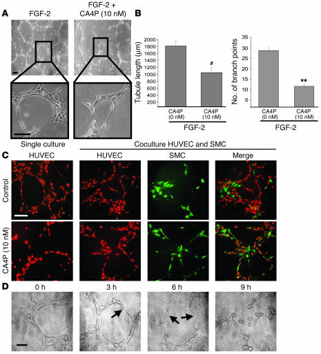
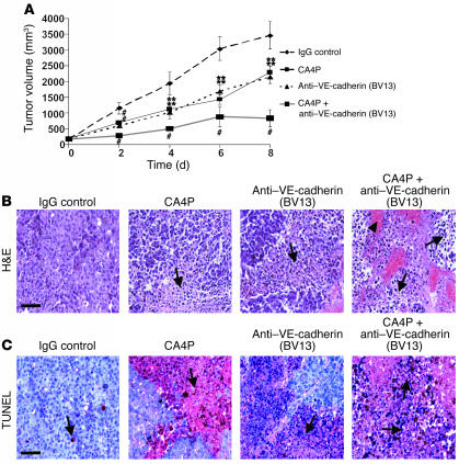
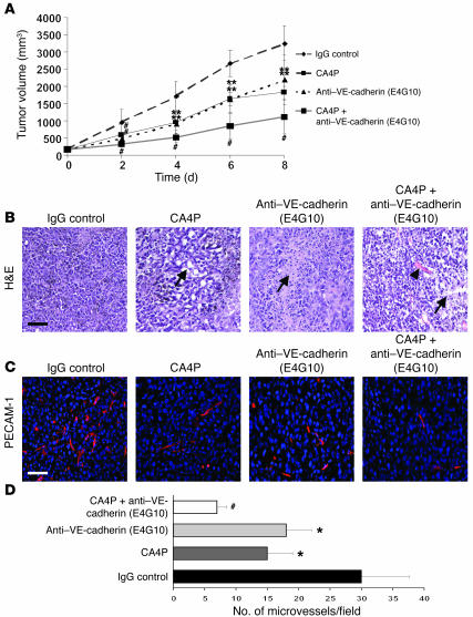
The molecular and cellular pathways that support the maintenance and stability of tumor neovessels are not well defined. The efficacy of microtubule-disrupting agents, such as combretastatin A4 phosphate (CA4P), in inducing rapid regression of specific subsets of tumor neovessels has opened up new avenues of research to identify factors that support tumor neoangiogenesis. Herein, we show that CA4P selectively targeted endothelial cells, but not smooth muscle cells, and induced regression of unstable nascent tumor neovessels by rapidly disrupting the molecular engagement of the endothelial cell-specific junctional molecule vascular endothelial-cadherin (VE-cadherin) in vitro and in vivo in mice. CA4P increases endothelial cell permeability, while inhibiting endothelial cell migration and capillary tube formation predominantly through disruption of VE-cadherin/beta-catenin/Akt signaling pathway, thereby leading to rapid vascular collapse and tumor necrosis. Remarkably, stabilization of VE-cadherin signaling in endothelial cells with adenovirus E4 gene or ensheathment with smooth muscle cells confers resistance to CA4P. CA4P synergizes with low and nontoxic doses of neutralizing mAbs to VE-cadherin by blocking assembly of neovessels, thereby inhibiting tumor growth. These data suggest that the microtubule-targeting agent CA4P selectively induces regression of unstable tumor neovessels, in part through disruption of VE-cadherin signaling. Combined treatment with anti-VE-cadherin agents in conjunction with microtubule-disrupting agents provides a novel synergistic strategy to selectively disrupt assembly and induce regression of nascent tumor neovessels, with minimal toxicity and without affecting normal stabilized vasculature.


Figures

Figure 1
Figure 1
CA4P inhibits growth factor–induced endothelial cell proliferation and migration. (A) CA4P inhibits HUVEC proliferation. HUVECs were incubated with or without growth factors, and CA4P was added at different concentrations. Cells were then counted at the indicated time points. Results of 4 experiments in duplicate are expressed as the mean number of cells ± SEM (*P < 0.05, **P < 0.01, #P < 0.001 compared with CA4P-untreated cells; n = 4). (B) SMCs are not sensitive to CA4P. HUVECs or SMCs were incubated with CA4P, and cells were counted after 48 hours. Results of 4 experiments in duplicate are expressed as the mean number of cells ± SEM (**P < 0.01, #P < 0.001 compared with CA4P-untreated cells; n = 4). (C) HUVECs are resistant to CA4P when cocultured with SMCs. HUVECs and SMCs were seeded together, incubated with CA4P, and counted after 48 hours. Results of 4 experiments in duplicate are expressed as the mean number of cells ± SEM. (D) CA4P inhibits HUVEC migration. A lesion was produced across the HUVECs’ monolayers, and unstimulated or FGF-2–stimulated cell monolayers were incubated with CA4P for 24 hours and then photographed. A representative picture is shown. Magnification: ×4. Scale bar, 500 μm. (E) Quantification of recovery of each denuded area after CA4P treatment. Results are expressed as the ratio of the number of invading cells to the number of migrating cells in absence of CA4P ± SEM (#P < 0.001 compared with CA4P-untreated cells; P < 0.001 compared with FGF-2–stimulated endothelial cells; n = 5).
Figure 2
Figure 2
CA4P impairs capillary tube formation, but has no effect on SMC-stabilized tubes, and destabilizes a preestablished vascular network. (A) CA4P inhibits capillary tube formation. HUVECs (3 × 10⁴) were seeded on Matrigel matrix and incubated in the presence of FGF-2 and with 10 nM CA4P. The effect of CA4P on capillary tube formation was observed after a 12-hour incubation under an inverted light microscope. Magnification: ×10 (top panels), ×40 (bottom panels). Scale bar, 10 μm (top panels), 15 μm (bottom panels). (B) Quantification of the CA4P-mediated capillary tube formation inhibition. The quantification was done by measuring the tubule length and counting the number of branch points in 4 different random pictures. Results are expressed as the mean of the tubule length and the mean of the number of branch points ± SEM (**P < 0.01, #P < 0.001 compared with CA4P-untreated cells; n = 4). (C) SMCs protect capillary tube formation against CA4P. HUVECs (3 × 10⁴) stained with red fluorescent cell linker and SMCs (1 × 10⁴) stained with green fluorescent cell linker were seeded together on Matrigel matrix and incubated in presence of FGF-2 and with 10 nM CA4P. The effect of CA4P on capillary tube formation was observed after a 12-hour incubation under an inverted fluorescent microscope. Magnification: ×20. Scale bar, 50 μm. (D) CA4P destabilizes a preestablished vascular network. HUVECs (3 × 10⁴) were seeded on Matrigel matrix and incubated in presence of FGF-2. Once the capillary network formed (after 12 hours), CA4P was added, and the effect of 10 nM CA4P on the disruption of the capillary network was monitored in a time-course manner every 3 hours. There was significant disruption of endothelial cell morphology (arrows). Magnification: ×40. Scale bar, 10 μm.
Figure 3
Figure 3
CA4P induces retraction of endothelial cells and disrupts actin cytoskeleton. (A) CA4P induces retraction of endothelial cells. The effect of CA4P (5 nM and 10 nM) on the cell shape of FGF-2–stimulated HUVECs was monitored during 48 hours by microscopy. Notice the retraction of HUVECs after 18 hours of treatment with CA4P. Magnification: ×20. Scale bar, 10 μm. (B) The actin cytoskeleton was stained with TRITC-labeled phalloidin and analyzed by fluorescent microscopy on FGF-2–stimulated HUVECs after an 18-hour incubation with CA4P. Addition of CA4P resulted in reorganization of actin stress fibers. Magnification: ×40. Scale bar, 5 μm.
Figure 4
Figure 4
CA4P blocks VE-cadherin/β-catenin/Akt signaling pathway. (A) CA4P disengages VE-cadherin and β-catenin. The localization of VE-cadherin and β-catenin in FGF-2–stimulated HUVECs was monitored during 18 hours by microscopy upon CA4P treatment (10 nM). Notice the redistribution of VE-cadherin and β-catenin assuming a zigzag pattern at cell-cell contacts (arrowheads) before the apparition of gaps between the cells (asterisks). Magnification: ×40. Scale bar, 10 μm. (B) Quantification of the organization pattern of VE-cadherin and β-catenin. The organization pattern, i.e., linear or zigzag, was evaluated by microscopic counting of 10 fields at ×10 magnification and presented as the percentage of linear or zigzag pattern per microscopic field ± SEM (#P < 0.001, P < 0.001 compared with linear and zigzag organization pattern observed at time 0, respectively; n = 10). (C) CA4P synergizes with human mAb against VE-cadherin to decrease cell viability. AdNullE4- or AdNullE4+-infected HUVECs were incubated with either CA4P or human mAb against VE-cadherin (BV9) or in combination, and the number of viable cells was determined after a 48-hour incubation. Results of 4 experiments in duplicate are expressed as the mean number of viable cells ± SEM (*P < 0.05, **P < 0.01, #P < 0.001 compared with untreated cells; n = 4). (D) CA4P synergizes with neutralizing mAb against VE-cadherin to increase endothelial cell permeability. Modification of endothelial cell permeability of AdNullE4- or AdNullE4+-infected HUVEC monolayers was assessed as described in Methods at designated time points. Results of 3 experiments in triplicate are expressed as the permeability ratio (experimental/control) ± SEM (*P < 0.05, **P < 0.01, #P < 0.001 compared with control; n = 3). (E) CA4P inhibits tyrosine phosphorylation of VE-cadherin and β-catenin. Levels of tyrosine phosphorylation of immunoprecipitated VE-cadherin and β-catenin were determined by Western blotting. (F) CA4P induces serine/threonine phosphorylation of β-catenin. Phosphorylation levels (Ser45 and Ser33/37/Thr41) of β-catenin were determined by Western blotting. (G) CA4P decreases Akt phosphorylation. Phosphorylation level (Ser 473) of Akt was determined by Western blotting. The condition where the serum-free medium (X-VIVO) was used corresponds to basal protein expression in the absence of stimulation.
Figure 5
Figure 5
Treatment with CA4P and neutralizing mAb against VE-cadherin (BV13) promotes B16 melanoma tumor necrosis. (A) Growth curves of IgG control–, CA4P-, anti–VE-cadherin– (BV13), or CA4P plus anti–VE-cadherin– (BV13) treated tumors. B16 melanoma cells were injected subcutaneously into the dorsa of C57BL/6 mice. Treatment was initiated after 10 days and mice injected every 2 days. Mice in the CA4P group received an i.v. injection of CA4P at 5 mg/kg; the anti–VE-cadherin group received an i.p. injection of 10 μg of mAb against VE-cadherin (BV13); the combined group received CA4P at 5 mg/kg plus 10 μg of mAb against VE-cadherin (BV13); and the control group received 10 μg of IgG antibody. Tumor size was measured until day 8 after the first injection, and tumor volumes were measured. The data represent the mean value of the tumor volume ± SEM (**P < 0.01, #P < 0.001 compared with IgG control group; n = 9). (B) Histopathological analysis of CA4P- and anti–VE-cadherin– (BV13) treated B16 melanoma tumors. Animals were sacrificed at day 8 after first injection. Shown here are H&E-stained paraffin sections demonstrating areas of necrosis (arrows) with CA4P and mAb against VE-cadherin (BV13) treatment. Arrowhead shows area of hemorrhage. Magnification: ×20. Scale bar, 80 μm. (C) Induction of tumor necrosis. Cell death within paraffin tumor sections was detected by TUNEL. Red staining represents positive signals within the tumors (blue cells are the negative, living cells). Arrows show areas of necrosis. Magnification: ×20. Scale bar, 80 μm.
Figure 6
Figure 6
CA4P synergizes with mAb against VE-cadherin (BV13) to block tumor neoangiogenesis. C57BL/6 mice bearing B16 melanoma cells were treated with IgG control, CA4P, anti–VE-cadherin (BV13), or CA4P plus anti–VE-cadherin (BV13) as described in Methods. After an 8-day treatment, tumors were removed and subjected to immunohistochemical analysis. (A) Vessel density determination in B16 melanoma tumors. The importance of intratumoral vascularization was assessed by PECAM-1 immunostaining (red fluorescence). Nuclei were detected by DAPI staining (blue). Note that numerous vessels are seen in the control group, whereas groups of mice treated with CA4P, anti–VE-cadherin (BV13), or combination have markedly fewer neovessels. Representative tumor sections of each group are shown. Magnification: ×20. Scale bar, 100 μm. (B) Quantification of the microvessel density in B16 melanoma tumor sections. The microvessel density is presented as mean number of microvessels per microscopic field ± SEM (*P < 0.05, #P < 0.001 compared with the IgG control group; n = 5). (C) CA4P impairs SMC-mediated stabilization of neovessels. The presence of endothelial cells and SMCs was assessed by fluorescence microscopy using PECAM-1 (red) and α-SMA (green) staining, respectively. Note that vessels in the CA4P and CA4P plus anti–VE-cadherin (BV13) groups are not positive for α-SMA, whereas in the absence of CA4P, vessels are surrounded by SMCs. Representative photographs of tumor sections are shown. Magnification: ×100. Scale bar, 10 μm. (D) Quantification of SMC-ensheathed vessels in B16 melanoma tumor sections. The number of SMC-ensheathed vessels is presented as mean number of SMC-ensheathed vessels per microscopic field ± SEM (#P < 0.001 compared with the IgG control group; n = 5).
Figure 7
Figure 7
CA4P synergizes with mAb against VE-cadherin (E4G10) to block tumor neoangiogenesis. C57BL/6 mice bearing B16 melanoma cells were treated with IgG control, CA4P, anti–VE-cadherin (E4G10), or CA4P plus anti–VE-cadherin (E4G10) as described in Methods. After an 8-day treatment, tumors were removed and subjected to immunohistochemical analysis. (A) Growth curves of IgG control–, CA4P-, anti–VE-cadherin– (E4G10), or CA4P plus anti–VE-cadherin– (E4G10) treated tumors. Tumor size was measured until day 8 after first injection. The data represent the mean value of the tumor volume ± SEM (**P < 0.01, #P < 0.001 compared with IgG control group; n = 9). (B) Histopathological analysis of CA4P- and anti–VE-cadherin– (E4G10) treated B16 melanoma tumors. Shown here are H&E-stained sections demonstrating areas of necrosis (arrows) with CA4P and mAb against VE-cadherin (E4G10) treatment. Arrowhead shows area of hemorrhage. Magnification: ×20. Scale bar, 80 μm. (C) Vessel density determination in B16 melanoma tumors. The importance of intratumoral vascularization was assessed by PECAM-1 immunostaining (red fluorescence). Nuclei were detected by DAPI staining (blue). Note that numerous vessels are seen in the control group, whereas groups of mice treated with CA4P, anti–VE-cadherin (E4G10), or combination have markedly fewer neovessels. Representative tumor sections of each group are shown. Magnification: ×20. Scale bar, 100 μm. (D) Quantification of the microvessel density in B16 melanoma tumor sections. The microvessel density is presented as mean number of microvessels per microscopic field ± SEM (*P < 0.05, #P < 0.001 compared with the IgG control group; n = 5).
Figure 8
Figure 8
CA4P used alone or in combination with mAb against VE-cadherin does not destabilize normal vasculature. (A) CA4P does not affect normal vasculature. Mouse ears were injected with CA4P (100 μg), mAb against VE-cadherin BV13 (10 μg), mAb against VE-cadherin E4G10 (150 μg), or a mix of CA4P plus mAb against VE-cadherin, and the control group received 150 μg of IgG antibody. Seven days after injection, ears were removed and stained for vessels using PECAM-1 staining. VIP-based immunodetection yielded a red reaction product. Magnification: ×20. (B) Quantitative analysis of the total vessel length in the mouse ears. Total vessel length was obtained from 5 peripheral fields for each ear of each animal (n = 5/group). Results show the total vessel length relative to that of control.
Figure 9
Figure 9
Proposed model for CA4P-mediated angiogenesis inhibition. Preexisting vessels are invested with SMCs protecting endothelial cells against CA4P-induced cell death. Because nascent unstable tumor neovessels are not ensheathed by periendothelial mural cells, CA4P selectively destabilizes neovessels by inducing VE-cadherin disengagement, therefore increasing the antiangiogenic effects of the neutralizing mAb against VE-cadherin without increasing toxicity to the normal vasculature.

Similar articles

Cited by

References

    1. Zhu Z, et al. Inhibition of human leukemia in an animal model with human antibodies directed against vascular endothelial growth factor receptor 2. Correlation between antibody affinity and biological activity. Leukemia. 2003;17:604–611. - PubMed
    1. Dejana E. Endothelial adherens junctions: implications in the control of vascular permeability and angiogenesis. J. Clin. Invest. 1996;98:1949–1953. - PMC - PubMed
    1. Breviario F, et al. Functional properties of human vascular endothelial cadherin (7B4/cadherin-5), an endothelium-specific cadherin. Arterioscler. Thromb. Vasc. Biol. 1995;15:1229–1239. - PubMed
    1. Carmeliet P, et al. Targeted deficiency or cytosolic truncation of the VE-cadherin gene in mice impairs VEGF-mediated endothelial survival and angiogenesis. Cell. 1999;98:147–157. - PubMed
    1. Caveda L, et al. Inhibition of cultured cell growth by vascular endothelial cadherin (cadherin-5/VE-cadherin) J. Clin. Invest. 1996;98:886–893. - PMC - PubMed

Publication types

MeSH terms