. 2006 Feb 15;394(Pt 1):237-42.
doi: 10.1042/BJ20051354.

Rescue of cell growth by sphingosine with disruption of lipid microdomain formation in Saccharomyces cerevisiae deficient in sphingolipid biosynthesis

Affiliations

Rescue of cell growth by sphingosine with disruption of lipid microdomain formation in Saccharomyces cerevisiae deficient in sphingolipid biosynthesis

Motohiro Tani et al. Biochem J. .

Abstract

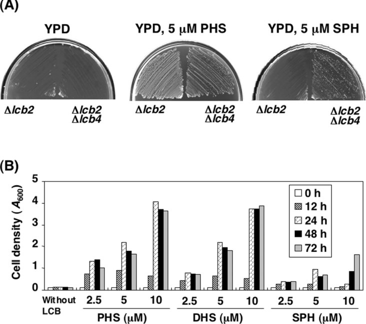
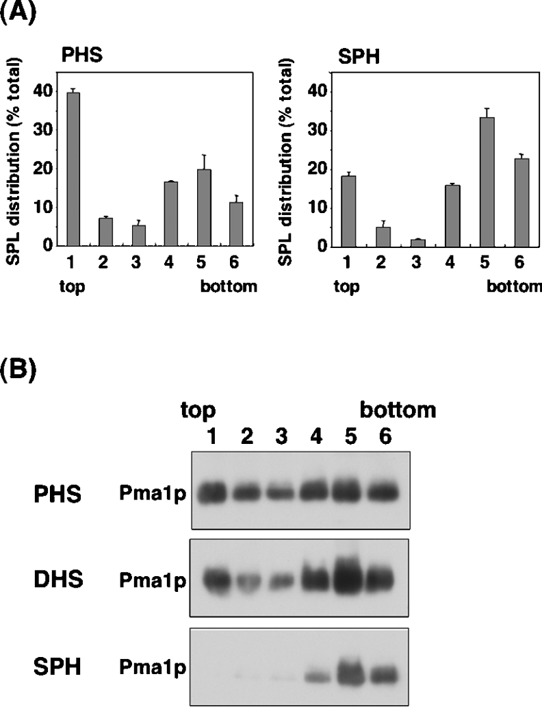
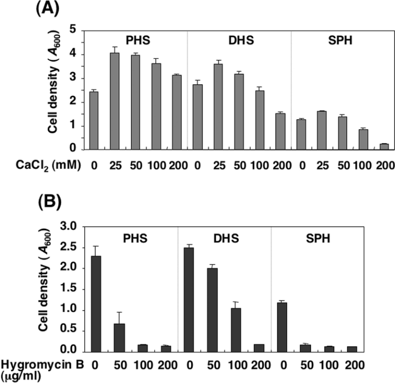
In the yeast Saccharomyces cerevisiae, sphingolipids are essential for cell growth. Inactivation of sphingolipid biosynthesis, such as by disrupting the serine palmitoyltransferase gene (LCB2), is lethal, but cells can be rescued by supplying an exogenous LCB (long-chain base) like PHS (phytosphingosine) or DHS (dihydrosphingosine). In the present study, supplying SPH (sphingosine), an unnatural LCB for yeast, similarly rescued the Deltalcb2 cells, but only when SPH 1-phosphate production was inhibited by deleting the LCB kinase gene LCB4. Exogenously added SPH was adequately converted into phosphoinositol-containing complex sphingolipids. Interestingly, cells carrying SPH-based sphingolipids exhibited a defect in the association of Pma1p with Triton X-100-insoluble membrane fractions, and displayed sensitivities to both Ca2+ and hygromycin B. These results suggest that the SPH-based sphingolipids in these cells have properties that differ from those of the PHS- or DHS-based sphingolipids in regard to lipid microdomain formation, leading to abnormal sensitivities towards certain environmental stresses. The present paper is the first report showing that in sphingolipid-deficient S. cerevisiae, the requirement for LCB can be fulfilled by exogenous SPH, although this supplement results in failure of lipid microdomain formation.


Figures

Figure 1
Figure 1. Deletion of LCB4 overcomes the growth defect of Δlcb2 cells cultured in the presence of SPH
(A) TMY77 (Δlcb2) and TMY60 (Δlcb2 Δlcb4) cells were streaked on to YPD plates in the absence or presence of 5 μM PHS or SPH and incubated for 3 days at 30 °C. (B) TMY85 (Δlcb2 Δlcb4 Δsur2) cells were cultured in YPD medium containing 5 μM PHS overnight. The cells were washed, diluted (0.1 D600 unit/ml) in YPD medium containing the indicated amounts of PHS, DHS or SPH and incubated at 30 °C. At the indicated times, the cell growth was determined by measuring the attenuance at 600 nm (D600) using a spectrophotometer. Results shown are the average for two independent experiments.
Figure 2
Figure 2. LCB analysis of LCB-reconstituted cells by HPLC
TMY85 cells were cultured overnight in YPD medium containing 5 μM PHS. The cells were washed and then diluted (0.1 D600 unit/ml) in YPD medium containing 5 μM PHS (B), 5 μM DHS (C) or 5 μM SPH (D), and incubated for 18 h at 30 °C. Lipids were extracted, hydrolysed by methanol/HCl, derivatized with OPA and analysed by reversed-phase HPLC. Details are provided in the Experimental section. (A) PHS, DHS and SPH standards.
Figure 3
Figure 3. Synthesis of complex sphingolipids in LCB-reconstituted cells
(A) [3H]myo-inositol labelling of LCB-reconstituted cells. TMY85 cells cultured with 5 μM PHS, DHS or SPH were labelled with [3H]myo-inositol for 5 h at 30 °C. Incorporated radioactivity was quantified, and equivalent samples (c.p.m.) were used for further analysis. Lipids were extracted and separated by TLC with chloroform/methanol/4.2 M ammonia (9:7:2, by vol.) as the solvent system. (B) Detection of unlabelled MIPCs in LCB-reconstituted cells. TMY85 cells (3.0 D600 units/ml) were cultured with PHS, DHS or SPH as in (A) and collected. Lipids were extracted, separated by TLC and then visualized by orcinol/H2SO4 reagent.
Figure 4
Figure 4. Effect of LCB reconstitution on the formation of lipid microdomains
TMY85 cells cultured with 5 μM PHS, DHS or SPH were lysed, incubated on ice with 1% Triton X-100 and fractionated by OptiPrep density-gradient centrifugation [49500 rev./min; SW60Ti (Beckman)]. Fractions were collected from the lowest-density (top) to the highest-density (bottom) gradient. (A) Distribution of sphingolipids (SPL) in PHS- or SPH-reconstituted cells. Lipids in each were hydrolysed by methanol/HCl. After derivatization with OPA, the hydrolysed LCBs were quantified by reversed-phase HPLC. Values presented are the amount of LCB in each fraction as a percentage of the total LCB in all fractions, and are the means±S.D. for three independent experiments. SPL, sphingolipids. (B) Association of Pma1p with the lipid microdomains. Fractions were subjected to SDS/PAGE, and Pma1p was detected by immunoblotting with an anti-Pma1p antibody. Details are provided in the Experimental section.
Figure 5
Figure 5. Effect of Ca2+ or the antibiotic hygromycin B on the growth of mutants deficient in sphingolipids cultured with various LCBs
TMY85 cells were cultured overnight in YPD medium containing 5 μM PHS. The cells were washed and diluted to 0.1 D600 unit/ml in YPD medium with 5 μM PHS, DHS or SPH and cultured at 30 °C for an additional 24 h in the absence or presence of increasing concentrations of CaCl2 (A) or hygromycin B (B). After a 24 h incubation, the cell growth was determined by measuring the attenuance at 600 nm (D600) using a spectrophotometer. Results shown are the means±S.D. for three independent experiments.

References

    1. Degroote S., Wolthoorn J., van Meer G. The cell biology of glycosphingolipids. Semin. Cell Dev. Biol. 2004;15:375–387. - PubMed
    1. Futerman A. H., Hannun Y. A. The complex life of simple sphingolipids. EMBO Rep. 2004;5:777–782. - PMC - PubMed
    1. Simons K., Ikonen E. Functional rafts in cell membranes. Nature (London) 1997;387:569–572. - PubMed
    1. Merrill A. H., Jr De novo sphingolipid biosynthesis: a necessary, but dangerous, pathway. J. Biol. Chem. 2002;277:25843–25846. - PubMed
    1. Dickson R. C., Lester R. L. Yeast sphingolipids. Biochim. Biophys. Acta. 1999;1426:347–357. - PubMed

Publication types

MeSH terms