Review
. 2005 Oct;23(4):351-60.
doi: 10.1016/j.immuni.2005.09.009.

Unusual features of self-peptide/MHC binding by autoimmune T cell receptors

Affiliations
Review

Unusual features of self-peptide/MHC binding by autoimmune T cell receptors

Melissa J Nicholson et al. Immunity. 2005 Oct.

Abstract

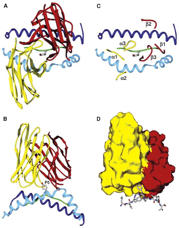
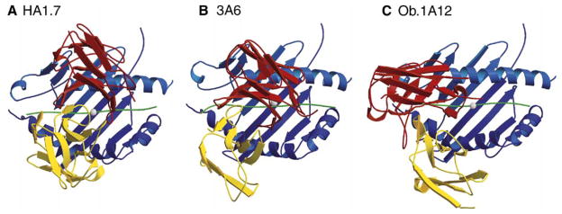
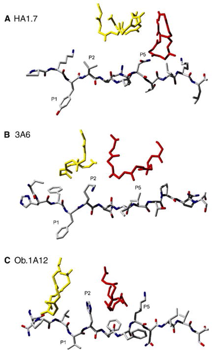
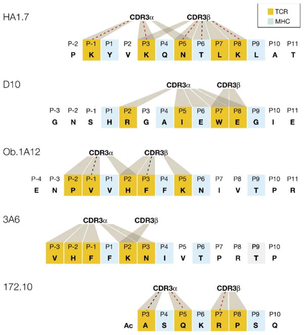
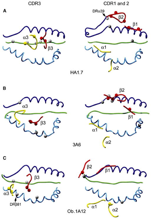
Structural studies on T cell receptors (TCRs) specific for foreign antigens demonstrated a remarkably similar topology characterized by a central, diagonal TCR binding mode that maximizes interactions with the MHC bound peptide. However, three recent structures involving autoimmune TCRs demonstrated unusual interactions with self-peptide/MHC complexes. Two TCRs from multiple sclerosis patients bind with unconventional topologies, and both TCRs are shifted toward the peptide N terminus and the MHC class II beta chain helix. A TCR from the experimental autoimmune encephalomyelitis (EAE) model binds in a conventional orientation, but the structure is unusual because the self-peptide only partially fills the binding site. For all three TCRs, interaction with the MHC bound self-peptide is suboptimal, and only two or three TCR loops contact the peptide. Optimal TCR binding modes confer a competitive advantage for antimicrobial T cells during an infection, whereas altered binding properties may permit survival of a subset of autoreactive T cells during thymic selection.


Figures

Figure 1
Figure 1. The Conventional Topology of TCR Binding to Peptide/MHC Complexes
(A and B) The conventional TCR binding mode is illustrated with the human A6 TCR as an example (Protein Data Bank [PDB] accession number 1AO7). This TCR recognizes the HTLV-1 Tax 11–19 peptide bound to HLA-A2 (Garboczi et al., 1996). The A6 TCR binds in a diagonal orientation to the peptide/MHC class I complex and is centered over the P5 tyrosine residue of the nine amino acid Tax peptide. Top view from the T cell surface (A) and side view (B) are shown; for clarity, only the TCR variable domains, the peptide backbone, and the MHC helices are shown. The side view shows the high points of the MHC helices that are avoided by the diagonal orientation of the TCR across the peptide/MHC surface. The P5 tyrosine side chain of the peptide is shown, and the backbone of the peptide is colored green. The MHC helices are colored blue (α1 helix in dark blue and α2 helix in light blue, respectively). The Vα and Vβ domains of TCR are colored yellow and red, respectively. The same color code is used throughout the figures for TCR V domains, MHC molecules, and peptides. (C) Location of the TCR loops on the peptide/MHC surface. The CDR3 loops of both TCR chains interact directly with the peptide and form a pocket for P5 tyrosine. The CDR1α and CDR1β loops are located over the N-terminal and C-terminal segments of the peptide, respectively. In the conventional diagonal orientation, the CDR2 loops do not contact the peptide, but make contacts to the MHC helices. The TCRα and TCRβ chain loops are colored yellow and red, respectively. The CDR1, CDR2, and CDR3 loops are labeled as α1, α2, and α3 for TCRα and as β1, β2, and β3 for TCRβ. (D) Space-filling model of the TCR variable domains (Vα yellow, Vβ red), illustrating the TCR pocket for the P5 tyrosine side chain of the peptide located in the center of the TCR interaction surface with peptide/MHC. The Tax peptide is shown as a stick-and-ball model. The figure was prepared with Deep View/Swiss-PDB Viewer (Guex and Peitsch, 1997).
Figure 2
Figure 2. Altered Topology of Self-Peptide/MHC Binding by Two Human Autoimmune TCRs
(A) The HA1.7 TCR (PDB accession number 1FYT) is specific for the influenza HA 306–318 peptide bound to DRA, DRB1*0101 and binds with a conventional topology over the center of the peptide/MHC surface. The white sphere marks the position of the P5 peptide residue in the center of the binding site over which the CDR3 loops of the TCRα and β chains converge. (B) The 3A6 TCR was isolated from a patient with MS and recognizes the MBP 89–101 peptide bound to DRA, DRB5*0101 (PDB accession number 1ZGL). Compared to HA1.7 TCR, the position of this TCR is shifted toward the peptide N terminus and the DRβ chain helix (the lower helix in all three DR molecules in this figure). (C) The Ob.1A12 TCR was isolated from another MS patient and recognizes a different epitope of MBP (residues 85–99) bound to another HLA-DR molecule (DRA, DRB1*1501) (PDB accession number 1YMM). This TCR is also shifted toward the peptide N terminus and tilted toward the DRβ chain helix. In addition, it is rotated counterclockwise relative to HA1.7 TCR. This counterclockwise rotation distinguishes it from 3A6 TCR. The TCR Vα and Vβ domains are colored in yellow and red, respectively, the MHC molecule in blue, and the bound peptide in green. The figure was prepared with Molscript (http://www.avatar.se/molscript/) and rendered with Raster3d (Merritt and Murphy, 1994).
Figure 3
Figure 3. Unusual Location of the TCR CDR3α and CDR3β Loops over the MHC Bound Peptide in the 3A6 and Ob.1A12 Structures
(A) The influenza HA peptide is shown as a stick model with the peptide backbone and side chain carbons in gray and the nitrogen and oxygen atoms in blue and red, respectively. The two CDR3 loops meet over the P5 side chain of the HA peptide. Only the backbone of the CDR3 loops (yellow, CDR3α; red, CDR3β) is shown to avoid crowding. (B and C) In the 3A6 (B) and Ob.1A12 (C) structures, the CDR3 loops are instead positioned over the P2 peptide residue, a lysine in the 3A6 structure, and a histidine in the Ob.1A12 structure. The CDR3β loop of Ob.1A12 TCR forms lateral contacts with the P5 lysine of the MBP peptide. The figure was prepared with Deep View/Swiss-PDB Viewer (Guex and Peitsch, 1997) and POV-Ray (http://www.povray.org/).
Figure 4
Figure 4. Peptide Contacts Established by TCR CDR3 Loops in Human and Murine TCR/Peptide/MHC Class II Structures
The peptide residues that occupy the P1, P4, P6, and P9 pockets of the MHC class II peptide binding site are colored light blue in all five peptide sequences. Peptide residues contacted by CDR3 loops are colored orange, and contacts to CDR3α or CDR3β are indicated by shaded areas. Contacts that represent hydrogen bonds are marked with a dotted line, which is colored red when the contact involves a side chain of the peptide. In the HA1.7 (human) and D10 (mouse) structures in which the TCR recognizes a foreign peptide, both CDR3 loops are located over the center of the peptide. In contrast, both human autoimmune TCRs (Ob.1A12 and 3A6) are characterized by a shift of the CDR3 loops toward the N terminus of the peptide. The mouse 172.10 TCR recognizes the N-terminal MBP Ac1-11 peptide that only partially fills the peptide binding site. This structure also contains a peptide extension by the insect leader peptide, which is not part of the native MBP peptide and thus not included in this figure. The CDR3 loops of D10 TCR do not form hydrogen bonds to the peptide, but a hydrogen bond is present between the CDR1α loop and P2 arginine of the peptide.
Figure 5
Figure 5. Location of the TCR Loops on the Peptide/MHC Surface in the Three Human TCR/Peptide/MHC Class II Structures
In the left panel, the CDR3 loops are located over the center of the peptide/MHC surface in the HA1.7 structure, but are shifted toward the peptide N terminus in the 3A6 and Ob.1A12 structures. The CDR3α (yellow) and CDR3β (red) loops are labeled as α3 and β3, respectively. Residues involved in hydrogen bonds are represented as small spheres. In the right panel, the CDR1 and CDR2 loops of TCRβ chains are located in different positions in each of the three structures, and their position is most extreme in the Ob.1A12 structure. The position of the CDR1 and CDR2 loops of TCRα chains is more similar among the three structures, but the CDR2α loop of 3A6 TCR fails to contact peptide/MHC. The CDR1 and CDR2 loops of TCRα and β are labeled as α1, α2, β1, and β2. The figure was prepared with Deep View/Swiss-PDB Viewer (Guex and Peitsch, 1997) and POV-Ray (http://www2.povray.org/).

References

    1. Alam SM, Travers PJ, Wung JL, Nasholds W, Redpath S, Jameson SC, Gascoigne NR. T-cell-receptor affinity and thymocyte positive selection. Nature. 1996;381:616–620. - PubMed
    1. Anderson AC, Nicholson LB, Legge KL, Turchin V, Zaghouani H, Kuchroo VK. High frequency of autoreactive myelin proteolipid protein-specific T cells in the periphery of naive mice: Mechanisms of selection of the self-reactive repertoire. J Exp Med. 2000;191:761–770. - PMC - PubMed
    1. Anderson MS, Venanzi ES, Klein L, Chen Z, Berzins SP, Turley SJ, von Boehmer H, Bronson R, Dierich A, Benoist C, Mathis D. Projection of an immunological self shadow within the thymus by the aire protein. Science. 2002;298:1395–1401. - PubMed
    1. Appel H, Gauthier L, Pyrdol J, Wucherpfennig KW. Kinetics of T-cell receptor binding by bivalent HLA-DR. Peptide complexes that activate antigen-specific human T-cells. J Biol Chem. 2000;275:312–321. - PubMed
    1. Baker BM, Turner RV, Gagnon SJ, Wiley DC, Biddison WE. Identification of a crucial energetic footprint on the alpha1 helix of human histocompatibility leukocyte antigen (HLA)-A2 that provides functional interactions for recognition by tax peptide/HLA-A2-specific T cell receptors. J Exp Med. 2001;193:551–562. - PMC - PubMed

Publication types

MeSH terms

Substances