. 2005 Nov;79(21):13209-17.
doi: 10.1128/JVI.79.21.13209-13217.2005.

Contribution of trafficking signals in the cytoplasmic tail of the infectious bronchitis virus spike protein to virus infection

Affiliations

Contribution of trafficking signals in the cytoplasmic tail of the infectious bronchitis virus spike protein to virus infection

Soonjeon Youn et al. J Virol. 2005 Nov.

Abstract

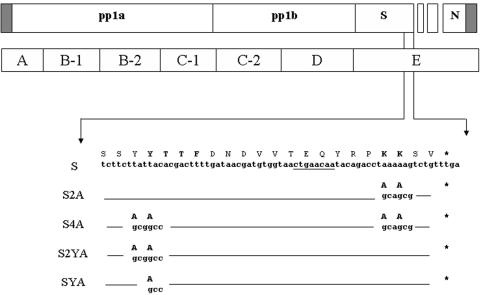
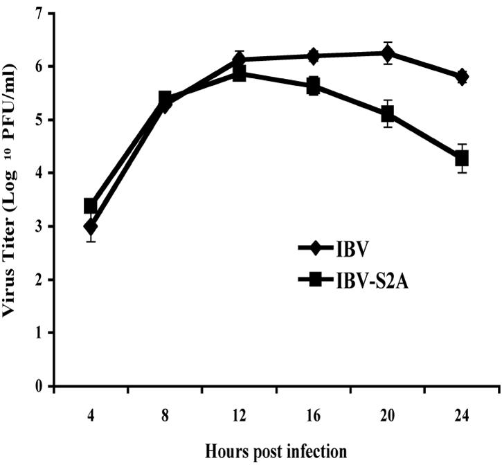
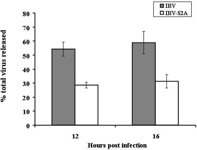
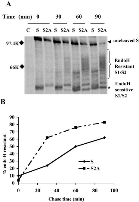
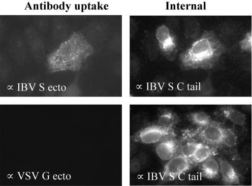
Coronavirus spike (S) proteins are responsible for binding and fusion with target cells and thus play an essential role in virus infection. Recently, we identified a dilysine endoplasmic reticulum (ER) retrieval signal and a tyrosine-based endocytosis signal in the cytoplasmic tail of the S protein of infectious bronchitis virus (IBV). Here, an infectious cDNA clone of IBV was used to address the importance of the S protein trafficking signals to virus infection. We constructed infectious cDNA clones lacking the ER retrieval signal, the endocytosis signal, or both. The virus lacking the ER retrieval signal was viable. However, this virus had a growth defect at late times postinfection and produced larger plaques than IBV. Further analysis confirmed that the mutant S protein trafficked though the secretory pathway faster than wild-type S protein. A more dramatic phenotype was obtained when the endocytosis signal was mutated. Recombinant viruses lacking the endocytosis signal (in combination with a mutated dilysine signal or alone) could not be recovered, even though transient syncytia were formed in transfected cells. Our results suggest that the endocytosis signal of IBV S is essential for productive virus infection.


Figures

FIG. 1.
FIG. 1.
Schematic diagram of IBV-S2A, IBV-S2YA, IBV-SYA, and IBV-S4A mutant virus constructs. The upper open boxes represent each major ORF of the IBV genome, and the closed boxes represent 5′ and 3′ untranslated regions. The lower boxes represent seven amplicons encompassing the entire genome of IBV used to construct recombinant IBVs. Amplicon E was used to eliminate the dilysine ER retrieval sequence located in the C terminus of the IBV S gene by replacing the codons for lysine with those for alanines, producing the S2A amplicon. The S4A amplicon was constructed using the S2A amplicon by additionally replacing the tyrosine codons for the endocytosis signal with those for alanines. The SYA and SY2A amplicons were constructed by replacing the single or double tyrosine codons in the endocytosis signal. Seven DNA fragments, including the E amplicon with or without mutations, were assembled by in vitro ligation and used to transcribe cRNAs. The partial nucleotide sequence and amino acid sequence of the IBV S C terminus are shown. The trafficking signals are shown in boldface. Lines indicate the same nucleotide and amino acid sequences as IBV. Asterisks represent stop codons. The underlined sequence represents the core TRS sequence of IBV subgenomic RNA3.
FIG. 2.
FIG. 2.
Defective growth of IBV-S2A at late times postinfection. One-step growth curves of IBV and IBV-S2A were performed. Vero cells were infected in triplicate with IBV or IBV-S2A at an MOI of 2. Total virus (supernatant plus cells disrupted by freeze-thaw) was harvested at the time points shown. Titers were measured by plaque assay at each time point. The error bars represent the standard errors of the means.
FIG. 3.
FIG. 3.
Recombinant IBV-S2A releases less infectious virus than IBV. Vero cells were infected in triplicate with IBV and IBV-S2A at an MOI of 2. The percentage of total virus released was determined after supernatants and cells were harvested separately at 12 and 16 h postinfection and titers were determined as described in Materials and Methods. The percentage of virus released was calculated by dividing the extracellular virus by the total (intracellular plus extracellular) virus. The error bars represent the standard errors of the means.
FIG. 4.
FIG. 4.
IBV-S2A produces larger plaques than IBV. (A) Vero cells were infected with IBV and IBV-S2A and overlaid with medium containing 0.8% agarose for 3 days. The agarose overlay was removed, and the cells were stained with crystal violet. Plaques produced by IBV are slightly smaller than plaques produced by IBV-S2A. (B) Average areas of plaques produced by IBV and IBV-S2A (IBV, n = 22; IBV-S2A, n = 30) were measured using Quantity One software (version 4.5.2). The error bars represent standard deviations. Student's t test, P = 0.00074.
FIG. 5.
FIG. 5.
S2A protein traffics through the Golgi faster than wild-type IBV S protein in infected cells. (A) At 8 h p.i., Vero cells infected with IBV and IBV-S2A were pulse-labeled with [35S]methionine-cysteine for 20 min and chased for the times indicated. Lysates were immunoprecipitated with anti-IBV S antibody and treated with endo H prior to sodium dodecyl sulfate-polyacrylamide gel electrophoresis. The arrowhead indicates endo H-sensitive uncleaved S0 protein, the bracket indicates the heterogeneous population of endo H-resistant cleaved S1/S2, and the asterisk indicates endo H-sensitive S1/S2. C, uninfected Vero cells; S, IBV-infected Vero cells; S2A, IBV-S2A-infected Vero cells. (B) The percentage of endo H-resistant S was calculated by dividing the amount of resistant S1/S2 by the total S for each time point. These data are representative of three independent experiments.
FIG. 6.
FIG. 6.
Mutation of the dilysine signal in IBV S affects transcription and translation of the downstream ORF. (A) At 24 h p.i., total cellular RNAs from wild-type IBV- or IBV-S2A-infected Vero cells were extracted and subjected to Northern blotting for subgenomic RNA profile analysis as described in Materials and Methods. Transcription of RNA3 (but not the other subgenomic RNAs) was significantly decreased in IBV-S2A-infected cells compared to that in cells infected with IBV. (B) E protein levels were analyzed by immunoprecipitation from [35S]methionine-cysteine-labeled infected Vero cells. At 9.5 h p.i., IBV- or IBV-S2A-infected Vero cells were labeled for 30 min and lysed, and the lysates were split in two and immunoprecipitated with anti-IBV E or anti-IBV M antibody. The results for two different clones of IBV-S2A (1 and 4) are shown.
FIG. 7.
FIG. 7.
Large transient syncytia formed after electroporation of cRNA with the S4A mutation, but no infectious virus could be recovered. In vitro-transcribed IBV or IBV-S4A cRNAs were electroporated into BHK-21 cells, which were then cocultured with Vero cells. (A) At 20 h posttransfection, normal-size syncytia started to form in IBV cRNA-transfected cells. (B) At 20 h posttransfection of IBV-S4A cRNA, transfected cells showed far fewer but much larger syncytia than IBV cRNA-transfected cells. (C) At 30 h posttransfection of IBV-S4A cRNA, the giant syncytia started to disappear by clumping and detaching from the dish. Uninfected Vero cells expanded to fill the empty areas left by the syncytia. Bars, 100 μm.
FIG. 8.
FIG. 8.
IBV-S4A interacts normally with IBV M. Interaction of S or S4A with IBV M protein was assessed by coimmunoprecipitation. (A) IBV S and M proteins interact in IBV-infected Vero cells. At 8 h p.i., Vero cells infected with IBV were labeled with [35S]methionine-cysteine for 30 min, and the cell lysate was subjected to immunoprecipitation with anti-IBV M or anti-IBV S antibody. (B) S or S4A protein was coexpressed with IBV M protein transiently using vaccinia virus expressing T7 RNA polymerase. After a 30-min pulse-labeling and 30-min chase in the presence or absence of BFA, S or S4A was coimmunoprecipitated with anti-IBV M antibody.
FIG. 9.
FIG. 9.
IBV S is rapidly endocytosed in infected cells. Vero cells on coverslips were infected with IBV at a MOI of 0.1 for approximately 18 h. A monoclonal antibody against the IBV S ectodomain (or the VSV G ectodomain as a negative control) was added to the culture medium for 15 min at 37°C. After being thoroughly washed, the cells were fixed, permeabilized, and stained for internal S protein using a rabbit antibody to the S cytoplasmic tail. Exogenously added monoclonal antibodies were detected using Texas red-conjugated goat anti-mouse IgG, and the internal antibody staining was detected with Alexa 488-conjugated goat anti-rabbit IgG. The left panels show internalized antibody, and the right panels show the internal staining of the same field. Note that only the cell with the higher intracellular level of IBV S endocytosed detectable antibody.

References

    1. Bonifacino, J. S., and L. M. Traub. 2003. Signals for sorting of transmembrane proteins to endosomes and lysosomes. Annu. Rev. Biochem. 72:395-447. - PubMed
    1. Corse, E., and C. E. Machamer. 2003. The cytoplasmic tails of infectious bronchitis virus E and M proteins mediate their interaction. Virology 312:25-34. - PMC - PubMed
    1. Corse, E., and C. E. Machamer. 2000. Infectious bronchitis virus E protein is targeted to the Golgi complex and directs release of virus-like particles. J. Virol. 74:4319-4326. - PMC - PubMed
    1. Danis, C., J. Deschambeault, S. Do Carmo, E. A. Cohen, E. Rassart, and G. Lemay. 2004. The tyrosine-based YXXO targeting motif of murine leukemia virus envelope glycoprotein affects pathogenesis. Virology 324:173-183. - PubMed
    1. de Haan, C. A., L. Kuo, P. S. Masters, H. Vennema, and P. J. Rottier. 1998. Coronavirus particle assembly: primary structure requirements of the membrane protein. J. Virol. 72:6838-6850. - PMC - PubMed

Publication types

MeSH terms

Substances

LinkOut - more resources