. 2005 Nov;79(21):13285-97.
doi: 10.1128/JVI.79.21.13285-13297.2005.

A major determinant for membrane protein interaction localizes to the carboxy-terminal domain of the mouse coronavirus nucleocapsid protein

Affiliations

A major determinant for membrane protein interaction localizes to the carboxy-terminal domain of the mouse coronavirus nucleocapsid protein

Kelley R Hurst et al. J Virol. 2005 Nov.

Abstract

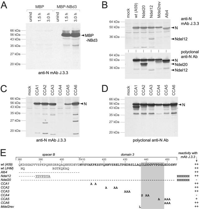
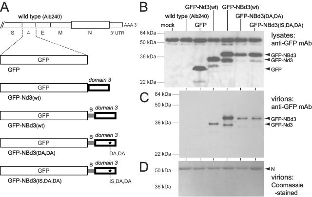
The two major constituents of coronavirus virions are the membrane (M) and nucleocapsid (N) proteins. The M protein is anchored in the viral envelope by three transmembrane segments flanked by a short amino-terminal ectodomain and a large carboxy-terminal endodomain. The M endodomain interacts with the viral nucleocapsid, which consists of the positive-strand RNA genome helically encapsidated by N protein monomers. In previous work with the coronavirus mouse hepatitis virus (MHV), a highly defective M protein mutant, MDelta2, was constructed. This mutant contained a 2-amino-acid carboxy-terminal truncation of the M protein. Analysis of second-site revertants of MDelta2 revealed mutations in the carboxy-terminal region of the N protein that compensated for the defect in the M protein. To seek further genetic evidence corroborating this interaction, we generated a comprehensive set of clustered charged-to-alanine mutants in the carboxy-terminal domain 3 of N protein. One of these mutants, CCA4, had a highly defective phenotype similar to that of MDelta2. Transfer of the CCA4 mutation into a partially diploid MHV genome showed that CCA4 was a loss-of-function mutation rather than a dominant-negative mutation. Analysis of multiple second-site revertants of CCA4 revealed mutations in both the M protein and the N protein that could compensate for the original lesion in N. These data more precisely define the region of the N protein that interacts with the M protein. Further, we found that fusion of domain 3 of the N protein to the carboxy terminus of a heterologous protein caused it to be incorporated into MHV virions.


Figures

FIG. 1.
FIG. 1.
Construction of a comprehensive set of CCA mutants in domain 3 of the N protein of MHV. (A) Schematic of the proposed structure of MHV N protein, with three domains separated by two short spacers (designated A and B) (22, 29, 39, 40). The expanded region of amino acid sequence shows the carboxy terminus of N protein, including part of spacer B and all of domain 3. Residue numbers and charged residues are indicated above the wild-type sequence; for CCA mutants 1 through 6, only those residues that differ from the wild type are shown. (B) Strategy for selection of mutant CCA4 by targeted RNA recombination between the interspecies chimera fMHV.v2 (17) and donor RNA transcribed from a derivative of plasmid pMH54 (23). fMHV.v2 contains the ectodomain-encoding region of the FIPV S gene (shaded rectangle) and grows in feline cells but not in murine cells. A single crossover within the HE gene should generate a recombinant that has simultaneously reacquired the MHV S ectodomain and the ability to grow in murine cells and has also incorporated the mutations in the N gene (star). The rearranged order of genes following the S gene in fMHV.v2 precludes the occurrence of a secondary crossover event downstream of the S gene (17). (C) Plaques of purified mutant CCA4 (Alb334) compared to the reconstructed wild type (Alb240) (25). Plaque titrations were carried out on mouse L2 cells at 37°C. Monolayers were stained with neutral red at 72 h postinfection and were photographed 18 h later.
FIG. 2.
FIG. 2.
Summary of the sequence analysis of the E, M, and N genes of revertants of CCA4 sibling isolates Alb334, Alb335, and Alb340. Revertants 1, 4 to 6, 10, 12, 13, 15, 25, and 26 were obtained from Alb334; revertants 2, 3, 7 to 9, 11, 14, 16, 27, and 28 were from Alb335; revertants 17 to 24 were from Alb340. The E, M, and N proteins are represented linearly at the top. Solid rectangles indicate the membrane-bound domains of the E and M proteins; the gray rectangle indicates the RNA-binding domain of the N protein (27, 35). Positions within the M and N proteins at which potential reverting mutations were found are connected by arrows to the wild-type residue, under which the changes found in each revertant are given. All coding changes are shown; the only other changes were silent mutations at codon 8 of the M protein and at codon 212 of the N protein of revertants 11 and 9, respectively. No changes were found in the E gene of any of the revertants. Boxed residues are those that were chosen for further analysis. At the bottom are shown the carboxy-terminal sequences of the N proteins of revertants 25 to 28 in comparison to those of the wild type and the CCA4 mutant. Amino acid residues that differ from those of the wild type, including insertions, are underlined; arrowheads indicate the positions of deletions.
FIG. 3.
FIG. 3.
Reconstruction of viruses containing the N gene adaptive mutation I438S and M gene reverting mutations of the CCA4 lesion. Derivative plasmids were constructed from the wild-type plasmid pSG6 (17), or from pSG6 containing the N gene CCA4 mutations (D440A and D441A) and the adaptive mutation I438S, in the presence or absence of individual candidate M gene reverting mutations. Donor RNAs transcribed from these vectors were used in targeted recombination experiments with fMHV.v2, as shown in the schematic in Fig. 1B, and progeny viruses were titrated directly on mouse L2 cells at 37°C. Monolayers were stained with neutral red at 72 h postinfection and were photographed 18 h later. In each case, plaques from two independent infection-transfections were subsequently purified by two rounds of plaque titration, and the presence of the expected mutant or wild-type sequences in the M and N genes was confirmed by direct sequencing of RT-PCR products prepared from RNA purified from virus-infected cells.
FIG. 4.
FIG. 4.
Rescue of the CCA4 mutant by wild-type N protein. (A) Schematic of the downstream end of the genome of the wild type (Alb240) and four recombinants in which the N gene is duplicated. In each, the wild-type copy of the N gene, in its normal genomic locus, is designated N[1]. In Alb284, the second copy of the N gene, designated N[2], is wild type. In Alb429 and Alb430, N[2] contains the CCA4 mutations D440A and D441A, and N[2] of Alb429 additionally contains the adaptive mutation I438S. In Alb330, N[2] contains a deletion of amino acids 134 to 248 within domain 2 of the molecule. The HA and His6 epitope tags are indicated by gray rectangles. (B) Plaques of the wild type (Alb240) and the N duplication mutants Alb284, Alb429, and Alb430. Plaque titrations were carried out on mouse L2 cells at 37°C. Monolayers were stained with neutral red at 72 h postinfection and were photographed 18 h later. (C to F) Western blots of lysates from cells infected with N duplication mutant Alb284, Alb330, Alb429, or Alb430 (C and D) or of purified virions (C, E, and F) probed with an antibody for the HA epitope tag (C), N protein (D and E), or M protein (F).
FIG. 5.
FIG. 5.
Localization of the epitope for anti-N MAb J.3.3 (15). (A) Western blots of lysates from bacterial cells that inducibly expressed either maltose binding protein (MBP) or maltose binding protein with a carboxy-terminal fusion of spacer B and domain 3 of the N protein (MBP-NBd3). Bacterial cultures were harvested prior to induction (unind) or after 1.5 or 3.0 h of induction. (B) Western blots of lysates from cells that were either mock infected or else infected with wild-type MHV-A59, N duplication mutant Ndel20 (Alb354) or Ndel12 (Alb381), or Mdel2rev (Alb302). The genomic arrangement of the N duplication mutants is similar to that of Alb330 (see Fig. 4). In Ndel20, the N[2] gene contains a deletion of amino acids 381 to 453 in spacer B and domain 3; in Ndel12, the N[2] gene contains a deletion of amino acids 188 to 387 in domain 2 and spacer B. Both Ndel20 and Ndel12 contain the His6 epitope tag; Ndel12 also retains a fragment of the HA epitope tag, while Ndel20 lacks it entirely. Mdel2rev is an intergenic revertant of the MΔ2 mutant that has the suppressor mutation Q437L in the N gene. (C and D) Western blots of lysates from cells that were mock infected or infected with CCA mutants 1 to 6. Blots were probed with MAb J.3.3 (A, top panel of B, and C) or polyclonal anti-N antibodies (bottom panel of B and D). (E) Summary of the abilities of various strains and mutants to cross-react with MAb J.3.3. The sequence of the carboxy terminus of the wild-type MHV-A59 N protein is shown at the top. For all other viruses, only residues that differ from the wild type (A59) are shown; dashes indicate deleted residues. In Alb4 and Ndel20, the entire deletion is shown. In Ndel12 only the downstream end of the deletion is represented, and the remnant of the HA tag is underlined. The gray rectangle indicates the region to which the MAb J.3.3 epitope maps.
FIG. 6.
FIG. 6.
Transfer of the M-interacting region of N protein to a heterologous protein. (A) Schematic of the downstream end of the genome of the wild type (Alb240) and five recombinants in which the GFP gene replaces the nonessential gene 4. Mutant GFP is identical to the MHV-A59 EGFP recombinant described previously (7). Mutant GFP-Nd3(wt) contains the wild-type domain 3 of the N protein linked to the carboxy terminus of GFP. Mutant GFP-NBd3(wt) contains the wild-type spacer B and domain 3 of the N protein. Mutants GFP-NBd3(DA,DA) and GFP-NBd3(IS,DA,DA) both contain spacer B and domain 3 with the CCA4 mutations D440A and D441A; GFP-NBd3(IS,DA,DA) additionally contains the adaptive mutation I438S. (B and C) Western blots of lysates from cells infected with the wild type or GFP-expressing mutants (B), or of purified virions (C), probed with an antibody for GFP. The amounts of virions analyzed in each lane of panel C were normalized to contain equivalent amounts of N protein, as determined by staining with Coomassie blue (D).
FIG. 7.
FIG. 7.
Genetic cross talk between the N and M proteins. The domain structure of N protein is shown linearly, as in Fig. 1. For the M protein, solid rectangles indicate the three transmembrane (tm) domains. Primary mutations that disrupt N-M interactions (the original lesions of the MΔ2 and CCA4 mutants) are boxed. The locations of second-site mutations, each of which individually suppresses one of the primary mutations, are indicated by lines or brackets; the asterisk represents a stop codon. The MΔ2 primary and reverting mutations (24) are in gray; the CCA4 primary and reverting mutations (this study) are in black. Arrows indicate compensating intramolecular and intermolecular interactions. (Inset) Schematic of the relative topologies and interfaces of the M, N, and E proteins at or within the virion envelope.

Similar articles

Cited by

References

    1. Baudoux, P., C. Carrat, L. Besnardeau, B. Charley, and H. Laude. 1998. Coronavirus pseudoparticles formed with recombinant M and E proteins induce alpha interferon synthesis by leukocytes. J. Virol. 72:8636-8643. - PMC - PubMed
    1. Bogan, A. A., and K. S. Thorn. 1998. Anatomy of hot spots in protein interfaces. J. Mol. Biol. 280:1-9. - PubMed
    1. Bos, E. C. W., W. Luytjes, H. van der Meulen, H. K. Koerten, and W. J. M. Spaan. 1996. The production of recombinant infectious DI-particles of a murine coronavirus in the absence of helper virus. Virology 218:52-60. - PMC - PubMed
    1. Corse, E., and C. E. Machamer. 2000. Infectious bronchitis virus E protein is targeted to the Golgi complex and directs release of virus-like particles. J. Virol. 74:4319-4326. - PMC - PubMed
    1. Corse, E., and C. E. Machamer. 2003. The cytoplasmic tails of infectious bronchitis virus E and M proteins mediate their interaction. Virology 312:25-34. - PMC - PubMed

Publication types

Substances

LinkOut - more resources