Amino acid residues within conserved domain VI of the vesicular stomatitis virus large polymerase protein essential for mRNA cap methyltransferase activity
- PMID: 16227259
- PMCID: PMC1262600
- DOI: 10.1128/JVI.79.21.13373-13384.2005
Amino acid residues within conserved domain VI of the vesicular stomatitis virus large polymerase protein essential for mRNA cap methyltransferase activity
Abstract
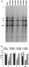
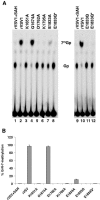
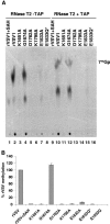
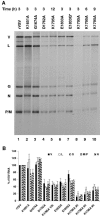
During mRNA synthesis, the polymerase of vesicular stomatitis virus (VSV) copies the genomic RNA to produce five capped and polyadenylated mRNAs with the 5'-terminal structure 7mGpppA(m)pApCpApGpNpNpApUpCp. The 5' mRNA processing events are poorly understood but presumably require triphosphatase, guanylyltransferase, [guanine-N-7]- and [ribose-2'-O]-methyltransferase (MTase) activities. Consistent with a role in mRNA methylation, conserved domain VI of the 241-kDa large (L) polymerase protein shares sequence homology with a bacterial [ribose-2'-O]-MTase, FtsJ/RrmJ. In this report, we generated six L gene mutations to test this homology. Individual substitutions to the predicted MTase active-site residues K1651, D1762, K1795, and E1833 yielded viruses with pinpoint plaque morphologies and 10- to 1,000-fold replication defects in single-step growth assays. Consistent with these defects, viral RNA and protein synthesis was diminished. In contrast, alteration of residue G1674 predicted to bind the methyl donor S-adenosylmethionine did not significantly perturb viral growth and gene expression. Analysis of the mRNA cap structure revealed that alterations to the predicted active site residues decreased [guanine-N-7]- and [ribose-2'-O]-MTase activity below the limit of detection of our assay. In contrast, the alanine substitution at G1674 had no apparent consequence. These data show that the predicted MTase active-site residues K1651, D1762, K1795, and E1833 within domain VI of the VSV L protein are essential for mRNA cap methylation. A model of mRNA processing consistent with these data is presented.
Figures


Similar articles
-
Identification of a new region in the vesicular stomatitis virus L polymerase protein which is essential for mRNA cap methylation.Virology. 2006 Jul 5;350(2):394-405. doi: 10.1016/j.virol.2006.02.021. Epub 2006 Mar 13. Virology. 2006. PMID: 16537083
-
A single amino acid change in the L-polymerase protein of vesicular stomatitis virus completely abolishes viral mRNA cap methylation.J Virol. 2005 Jun;79(12):7327-37. doi: 10.1128/JVI.79.12.7327-7337.2005. J Virol. 2005. PMID: 15919887 Free PMC article.
-
Analysis of a structural homology model of the 2'-O-ribose methyltransferase domain within the vesicular stomatitis virus L protein.Virology. 2008 Dec 5;382(1):69-82. doi: 10.1016/j.virol.2008.08.041. Epub 2008 Oct 11. Virology. 2008. PMID: 18848710 Free PMC article.
-
An unconventional pathway of mRNA cap formation by vesiculoviruses.Virus Res. 2011 Dec;162(1-2):100-9. doi: 10.1016/j.virusres.2011.09.012. Epub 2011 Sep 16. Virus Res. 2011. PMID: 21945214 Free PMC article. Review.
-
[The multifunctional RNA polymerase L protein of non-segmented negative strand RNA viruses catalyzes unique mRNA capping].Uirusu. 2014;64(2):165-78. doi: 10.2222/jsv.64.165. Uirusu. 2014. PMID: 26437839 Review. Japanese.
Cited by
-
Molecular architecture of the vesicular stomatitis virus RNA polymerase.Proc Natl Acad Sci U S A. 2010 Nov 16;107(46):20075-80. doi: 10.1073/pnas.1013559107. Epub 2010 Nov 1. Proc Natl Acad Sci U S A. 2010. PMID: 21041632 Free PMC article.
-
A Zika virus vaccine expressing premembrane-envelope-NS1 polyprotein.Nat Commun. 2018 Aug 3;9(1):3067. doi: 10.1038/s41467-018-05276-4. Nat Commun. 2018. PMID: 30076287 Free PMC article.
-
Inhibition of viral RNA-dependent RNA polymerases with clinically relevant nucleotide analogs.Enzymes. 2021;49:315-354. doi: 10.1016/bs.enz.2021.07.002. Epub 2021 Oct 15. Enzymes. 2021. PMID: 34696837 Free PMC article.
-
Lack of correlation between virus barosensitivity and the presence of a viral envelope during inactivation of human rotavirus, vesicular stomatitis virus, and avian metapneumovirus by high-pressure processing.Appl Environ Microbiol. 2011 Dec;77(24):8538-47. doi: 10.1128/AEM.06711-11. Epub 2011 Oct 14. Appl Environ Microbiol. 2011. PMID: 22003028 Free PMC article.
-
N6-methyladenosine modification enables viral RNA to escape recognition by RNA sensor RIG-I.Nat Microbiol. 2020 Apr;5(4):584-598. doi: 10.1038/s41564-019-0653-9. Epub 2020 Feb 3. Nat Microbiol. 2020. PMID: 32015498 Free PMC article.
References
-
- Abraham, G., D. P. Rhodes, and A. K. Banerjee. 1975. The 5′ terminal structure of the methylated mRNA synthesized in vitro by vesicular stomatitis virus. Cell 5:51-58. - PubMed
Publication types
MeSH terms
Substances
Grants and funding
LinkOut - more resources
Full Text Sources
Other Literature Sources
Miscellaneous

