. 2005 Nov;79(21):13385-98.
doi: 10.1128/JVI.79.21.13385-13398.2005.

Kelp fly virus: a novel group of insect picorna-like viruses as defined by genome sequence analysis and a distinctive virion structure

Affiliations

Kelp fly virus: a novel group of insect picorna-like viruses as defined by genome sequence analysis and a distinctive virion structure

C J Hartley et al. J Virol. 2005 Nov.

Abstract

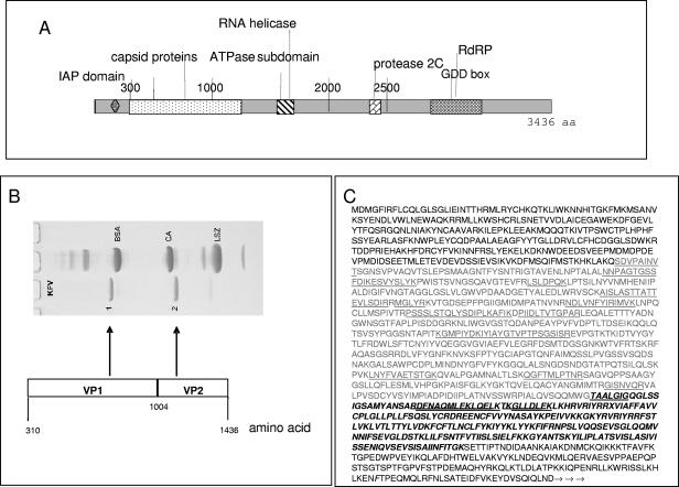
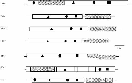
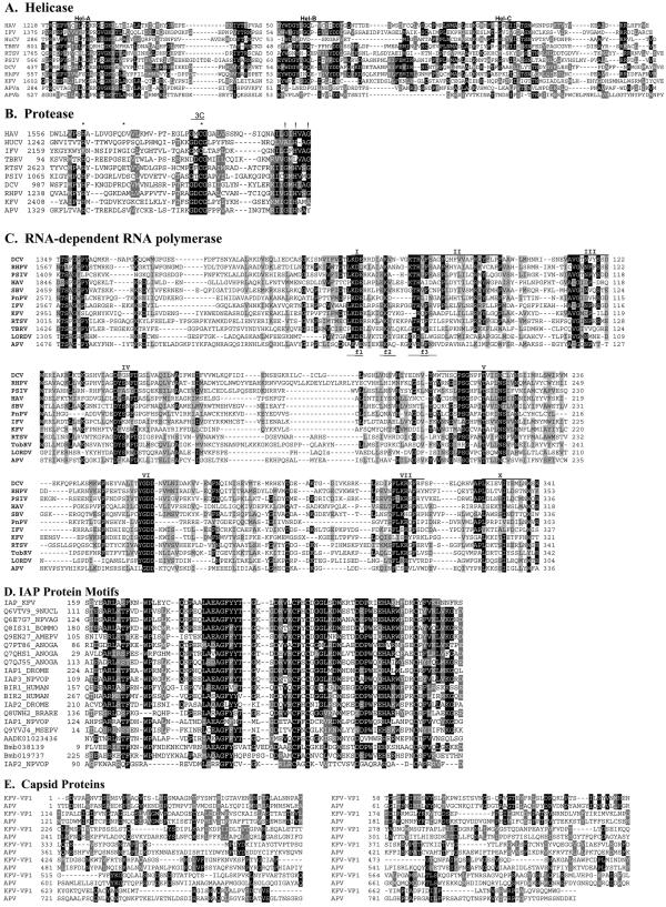
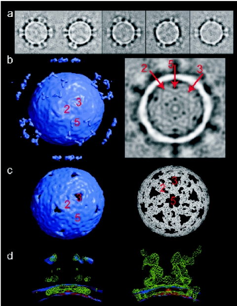
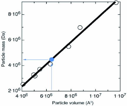
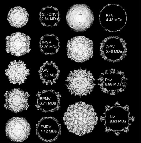
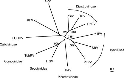
The complete genomic sequence of kelp fly virus (KFV), originally isolated from the kelp fly, Chaetocoelopa sydneyensis, has been determined. Analyses of its genomic and structural organization and phylogeny show that it belongs to a hitherto undescribed group within the picorna-like virus superfamily. The single-stranded genomic RNA of KFV is 11,035 nucleotides in length and contains a single large open reading frame encoding a polypeptide of 3,436 amino acids with 5' and 3' untranslated regions of 384 and 343 nucleotides, respectively. The predicted amino acid sequence of the polypeptide shows that it has three regions. The N-terminal region contains sequences homologous to the baculoviral inhibitor of apoptosis repeat domain, an inhibitor of apoptosis commonly found in animals and in viruses with double-stranded DNA genomes. The second region contains at least two capsid proteins. The third region has three sequence motifs characteristic of replicase proteins of many plant and animal viruses, including a helicase, a 3C chymotrypsin-like protease, and an RNA-dependent RNA polymerase. Phylogenetic analysis of the replicase motifs shows that KFV forms a distinct and distant taxon within the picorna-like virus superfamily. Cryoelectron microscopy and image reconstruction of KFV to a resolution of 15 A reveals an icosahedral structure, with each of its 12 fivefold vertices forming a turret from the otherwise smooth surface of the 20-A-thick capsid. The architecture of the KFV capsid is unique among the members of the picornavirus superfamily for which structures have previously been determined.


Figures

FIG. 1.
FIG. 1.
KFV genome. (A) Graphical illustration showing the peptide domains within the full-length KFV polypeptide coding sequence and UTRs. aa, amino acid. (B) Coomassie-stained SDS-PAGE illustrating the structural proteins isolated from KFV virions and the corresponding coding regions within the KFV genome. BSA, bovine serum albumin; CA, carbonic anhydrase; LSZ, lysozyme. (C) Virus capsid protein sequence. The inferred amino acid sequence begins at the start of the single KFV ORF. The light gray type indicates VP1 residues, and the bold and italic type represents the probable extent of VP2 residues. The N-terminal sequences SDVPAINVT and TAALGIG were determined as described in Materials and Methods. These and other sequences determined by mass spectrometry of tryptic digests of KFV capsid proteins are underlined.
FIG. 2.
FIG. 2.
Genomic arrangement of KFV peptide domains within the full-length KFV polypeptide sequence and comparison to other picorna-like insect viruses. Lines represent UTRs, and boxes represent ORFs. Shaded areas represent capsid proteins, and objects within the boxes represent the following: RdRp (▪), protease (•), RNA helicase (▴), and IAP domain (⧫). DCV, Drosophila C virus; RhPV, Rhopalosiphum padi virus; PSIV, Plautia stali intestine virus.
FIG. 3.
FIG. 3.
Comparison of the deduced amino acid sequences of the structural and nonstructural proteins of KFV and other picorna-like viruses. (A) Alignment of the conserved regions of the putative RNA helicase protein sequence from KFV with those of other picorna-like virus. The motifs recognized by Gorbalenya et al. (27) are labeled Hel-A, Hel-B, and Hel-C. (B) Alignment of KFV and picorna-like RNA virus chymotrypsin-related cysteine protease 3C-like protein sequences. The conserved residues of chymotrypsin-like proteases (26) that should form the catalytic triad are marked with asterisks, and putative substrate-binding residues are indicated by exclamation marks. The conserved catalytic cysteine motif associated with 3C proteases is indicated by the region labeled “3C.” (C) Alignment of KFV and picorna-like virus RdRp protein sequences. The amino acid sequences were aligned using the GAP program as described by Gorbalenya et al. (28). The motifs reviewed by Koonin and Dolja (36) are labeled I to VII. The f1 to f3 regions of the conserved motif F recognized by Bruenn (4) are denoted as such below the alignment sequences. (D) Comparison of the deduced amino acid sequences of representative BIR proteins of KFV, viral, dipteran, lepidopteran, and vertebrate origins. The KFV domain is labeled IAP_KFV. Other amino acid sequences are from UniProt (labeled with their entry numbers). The only exceptions are three possible novel IAPs from the Bombyx mori genome (64); these are predicted proteins Bmb038139 and Bmb019737, plus a partial sequence present in contig AADK01023436. Sequence identities of the sequences shown to the KFV sequence range from 58% identity (E = 7e−22) for the Choristoneura fumiferana defective nucleopolyhedrovirus (Q6VTV9_9NUCL) at the top down to ca. 30% for the IAP2 from the Orgyia pseudotsugata multicapsid polyhedrosis virus (IAP2_NPVOP). (E) Comparison of the deduced amino acid sequences of the structural proteins of KFV (VP1) and APV. Numbers to the initial left of alignments represent the residue numbers within the GenBank sequences. Numbers thereafter refer to residue distance from that point. Black shading indicates ≥50% identity; gray shading indicates ≥50% similarity. DCV, Drosophila C virus; RhPV, Rhopalosiphum padi virus; PSIV, Plautia stali intestine virus; PnPV, Perina nuda picorna-likevirus; HAV, human hepatitis A virus; RTSV, rice tungro spherical virus; HuCV, human calicivirus; TBRV and TobRV, tobacco ringspot virus.
FIG. 4.
FIG. 4.
Three-dimensional reconstruction of KFV by cryo-EM. (a) Gallery of class averages of raw KFV images, ranked by turret occupancy. This shows that there is no change in overall capsid morphology with the loss of turrets and that the turret structure is the same when fully occupied as when partially occupied. (b) Left: surface-rendered view (contour 2σ) of an icosahedral reconstruction of KFV. Right: central section through the same map. The symmetry axes are marked in both cases. (c) Left: surface-rendered view (contour 4σ) of the map shown in panel a. Right: the same map in another view at the 5.2σ-contour level. (d) Superpositions of a reconstruction selected for low turret occupancy (red), the reconstruction shown in panels b and c (lilac), and a difference map (green), viewed across a fivefold vertex. The difference map highlights features present in the partially turreted map but absent from the map constructed from particles selected for the absence of turrets. Left: reconstructions at 2σ and difference map at 3σ. Right: these thresholds are switched.
FIG. 5.
FIG. 5.
Plot of the atomic mass of virus particles (capsid shell) against the volume of low-resolution maps calculated from their atomic coordinates. The lilac plot point marks the positioning of the KFV shell in this range of masses, with the lilac arrows showing that a volume of 6.41 × 10⁶ Å3 gives the KFV shell a mass of 4.48 MDa.
FIG. 6.
FIG. 6.
A gallery of views of virus structures, including insect members of the picornavirus superfamily, definitive picornaviruses, and other insect viruses. In each case, the rendered surface of the structure (at an 18-Å resolution) is shown, as well as a central section through the map. The masses marked are those of the atomic structures shown at lower-resolution density here, except for KFV, which is a mass value based on its capsid surface volume normalized to the mass/volume ratio of the other structures (see Fig. 5). The viruses shown are Galleria mellonella densovirus (GmDNV) (a parvovirus with a T=1 capsid; total genome size, 5 kb [53]); tobacco ringspot virus (TRSV) (a nepovirus with one coat protein making a pseudo T=3 capsid; total genome size, 15.5 kb [8]); adeno-associated virus (AAV) (with a T=1 capsid [65]); bean pod mottle virus (BPMV) (a comovirus with two coat proteins making a pseudo T=3 capsid; total genome size, 9.7 kb [41]); FMDV (a classical picornavirus with a pseudo T=3 capsid; total genome size, 8.4 kb [1]); KFV (total genome size, 11 kb); CrPV (a pseudo T=3 virus; total genome size, 9.2 kb [56]); Pariacoto virus (PaV) (a nodavirus with a T=3 capsid; total genome size, 4.3 kb [55]); and Norwalk virus (NV) (a calicivirus with a proper T=3 capsid; total genome size, 7.9 kb [9, 49]).
FIG. 7.
FIG. 7.
Resolving the KFV turret structure. (a) Views of the reconstruction of a single-turreted KFV, viewed from above the virus surface (left, surface not in view) and from the side, through the fivefold vertex (right). The map is contoured at three levels, 1.5σ, 2σ, and 4σ, to show the relative strengths of the density for different portions of the turret and the steep density gradient defining the edges of the structure. Identical features in the two views are labeled i to iii. (b) Views down fivefold, threefold, and twofold axes (left to right, respectively) of the complete KFV capsid generated using icosahedral symmetry from an asymmetric unit derived from the single-turreted reconstruction.
FIG. 8.
FIG. 8.
Phylogenetic analysis of the relationship of KFV RdRp sequences to those of other virus families and viruses in the picornavirus superfamily. The phenogram is based on the alignment in Fig. 3C of polymerases from KFV and representative viruses of the Picornaviridae, Dicistroviridae, Comoviridae, Sequiviridae, and Caliciviridae families, as well as the unclassified insect virus APV. An unrooted neighbor-joining tree was inferred using the programs ClustalX 1.81 (58) and PHYLIP (19). All bifurcations with support in >700 out of 1,000 bootstraps are indicated. DCV, Drosophila C virus; RhPV, Rhopalosiphum padi virus; PSIV, Plautia stali intestine virus; PnPV, Perina nuda picorna-like virus; HAV, human hepatitis A virus; RTSV, rice tungro spherical virus; TobRV, tobacco ringspot virus.

References

    1. Acharya, R., E. E. Fry, D. I. Stuart, G. Fox, D. J. Rowlands, and F. Brown. 1989. The three-dimensional structure of foot-and-mouth disease virus at 2.9 Å resolution. Nature 337:709-716. - PubMed
    1. Altschul, S. F., T. L. Madden, A. A. Schaffer, J. Zhang, Z. Zhang, W. Miller, and D. J. Lipman. 1997. Gapped BLAST and PSI-BLAST: a new generation of protein database search programs. Nucleic Acids Res. 25:3389-3402. - PMC - PubMed
    1. Birnbaum, M. J., R. J. Clem, and L. K. Miller. 1994. An apoptosis-inhibiting gene from a nuclear polyhedrosis virus encoding a polypeptide with Cys/His sequence motifs. J. Virol. 68:2521-2528. - PMC - PubMed
    1. Bruenn, J. A. 2003. A structural and primary sequence comparison of the viral RNA-dependent RNA polymerases. Nucleic Acids Res. 31:1821-1829. - PMC - PubMed
    1. Bruenn, J. A. 1991. Relationships among the positive strand and double-strand RNA viruses as viewed through their RNA-dependent RNA polymerases. Nucleic Acids Res. 19:217-226. - PMC - PubMed

Publication types

Substances

Associated data

LinkOut - more resources