. 2005 Nov;79(21):13421-33.
doi: 10.1128/JVI.79.21.13421-13433.2005.

VP24 of Marburg virus influences formation of infectious particles

Affiliations

VP24 of Marburg virus influences formation of infectious particles

Sandra Bamberg et al. J Virol. 2005 Nov.

Abstract

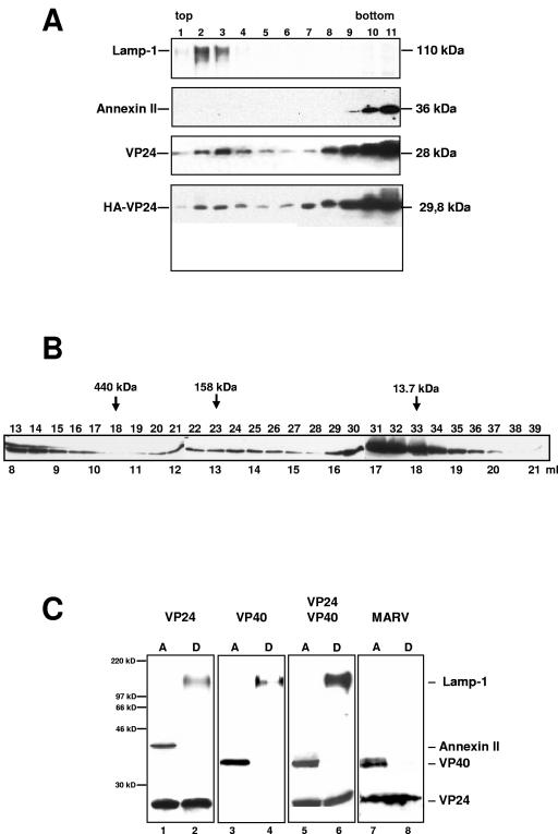
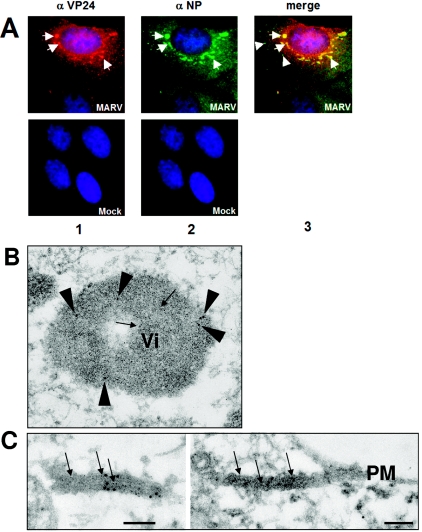
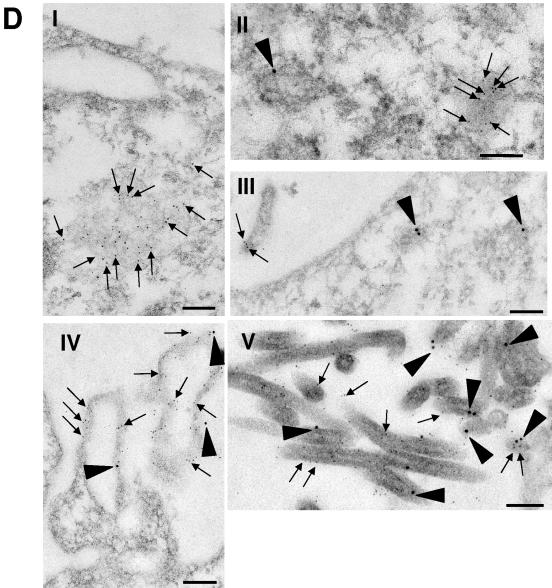
The highly pathogenic enveloped Marburg virus (MARV) is composed of seven structural proteins and the nonsegmented negative-sense viral RNA genome. Four proteins (NP, VP35, VP30, and L) make up the helical nucleocapsid, which is surrounded by a matrix that is composed of the viral proteins VP40 and VP24. VP40 is functionally homologous to the matrix proteins of other nonsegmented negative-strand RNA viruses. As yet, the function of VP24 remains elusive. In the present study we found that VP24 colocalized with inclusions in MARV-infected cells that contain preformed nucleocapsids and with nucleocapsids outside the inclusions. Coexpression studies revealed that VP24 is recruited into the inclusions by the presence of NP. Furthermore, VP24 displayed membrane-binding properties and was recruited into filamentous virus-like particles (VLPs) that are induced by VP40. The incorporation of VP24 altered neither the morphology of VLPs nor the budding efficiency of VLPs. When VP24 was silenced in MARV-infected cells by small interfering RNA technology, the release of viral particles was significantly reduced while viral transcription and replication were unimpaired. Our data support the idea that VP24 is essential for a process that takes place after replication and transcription and before budding of virus progeny. It is presumed that VP24 is necessary for the formation of transport-competent nucleocapsids and/or the interaction between the nucleocapsids and the budding sites at the plasma membrane.


Figures

FIG. 1.
FIG. 1.
Membrane association of VP24. (A) HEK293 cells were transfected with pC-VP24 and harvested at 24 h posttransfection, and cell lysates were subjected to flotation analysis via discontinuous sucrose gradient. The gradient was centrifuged to equilibrium at 35,000 rpm overnight in a Beckman SW41 rotor. Fractions were collected from the top, and samples were analyzed by SDS-PAGE and Western blotting. Membranes were stained with anti-LAMP-1 (monoclonal), anti-annexin II (monoclonal), and anti-VP24 (rabbit), respectively. Top of the gradient, fraction 1; bottom of the gradient, fraction 11. (B) Gel filtration and Western blot analyses of VP24. HEK293 cells were transfected with pC-VP24, and cell extracts were harvested at 36 h posttransfection. Clarified and filtered cell lysate was separated by size on a Superdex-200 10/30 high-resolution fast-performance liquid chromatography column. Molecular mass standards were also separated on the same column. A Western blot analysis of fractions representing elution volumes of 8.0 to 21.0 ml is shown. One peak of VP24 eluted within the 13.5-ml volume, which corresponds to a protein of approximately 120 kDa (tetramer). The majority of VP24 appeared at elution volume of 17.5 ml and corresponded to the monomeric form of VP24 (∼28 kDa). (C) Triton X-114 phase partitioning analysis of VP24. HEK293 cells were transfected with pC-VP24, pC-VP40, or both plasmids and cells were harvested at 24 h posttransfection. MARV particles from the supernatant of infected Vero cells were purified over a sucrose cushion prior to phase partitioning. Postnuclear supernatants of HEK293 cells and purified MARV particles were partitioned into aqueous (A) and detergent (D) phases as described in Materials and Methods and analyzed by SDS-PAGE and Western blotting. Blots were cut horizontally to get pieces corresponding to the targeted proteins, and the pieces were stained separately using anti-LAMP-1 (mouse monoclonal), anti-annexin II (mouse monoclonal), anti-VP24 (rabbit), and anti-VP40 (rabbit) antibodies, respectively.
FIG.2.
FIG.2.
Immunofluorescence and immunoelectron microscopic analysis of VP24 in MARV-infected cells. (A) Distribution of VP24 in MARV-infected cells. Vero cells were infected with MARV and fixed 24 h postinfection. For immunofluorescence analysis, a rabbit anti-VP24 antibody and a monoclonal anti-NP antibody were used as primary antibodies. Donkey anti-rabbit immunoglobulin G conjugated with rhodamine and goat anti-mouse antibody conjugated with FITC were used as secondary antibodies. Arrows, colocalization of VP24 with NP; arrowheads, singly located NP. (B to D) Immunoelectron microscopic analysis of the ultrathin sections of MARV-infected Vero cells. Vero cells were fixed at 48 h postinfection and embedded in LR Gold, and ultrathin sections were subjected to immunoelectron microscopy. (B and C) Samples were stained using rabbit anti-VP24 and mouse anti-NP primary antibodies. Secondary antibodies were used as described for panel D. Arrows, NP; arrowheads, VP24. Bar, 100 nm. (B) Viral inclusion (Vi). (C) Single nucleocapsid in the cytoplasm (left) and single nucleocapsid near the plasma membrane (PM, right). (D) Samples were stained using rabbit anti-VP24 and mouse anti-VP40 primary antibodies. As secondary antibodies goat anti-rabbit antibody conjugated with 12-nm gold and goat anti-mouse antibody conjugated with 6-nm gold were used. Arrows, VP40; arrowheads, VP24. Bar, 200 nm. Shown are a multivesicular body containing VP40 (I), cytoplasm containing separately located VP40 and VP24 (II), cellular protrusion with VP40 and VP24 below the plasma membrane (III), cellular protrusions containing both VP24 and VP40 at the plasma membrane (IV), and released viral particles containing VP40 and VP24 (V).
FIG.2.
FIG.2.
Immunofluorescence and immunoelectron microscopic analysis of VP24 in MARV-infected cells. (A) Distribution of VP24 in MARV-infected cells. Vero cells were infected with MARV and fixed 24 h postinfection. For immunofluorescence analysis, a rabbit anti-VP24 antibody and a monoclonal anti-NP antibody were used as primary antibodies. Donkey anti-rabbit immunoglobulin G conjugated with rhodamine and goat anti-mouse antibody conjugated with FITC were used as secondary antibodies. Arrows, colocalization of VP24 with NP; arrowheads, singly located NP. (B to D) Immunoelectron microscopic analysis of the ultrathin sections of MARV-infected Vero cells. Vero cells were fixed at 48 h postinfection and embedded in LR Gold, and ultrathin sections were subjected to immunoelectron microscopy. (B and C) Samples were stained using rabbit anti-VP24 and mouse anti-NP primary antibodies. Secondary antibodies were used as described for panel D. Arrows, NP; arrowheads, VP24. Bar, 100 nm. (B) Viral inclusion (Vi). (C) Single nucleocapsid in the cytoplasm (left) and single nucleocapsid near the plasma membrane (PM, right). (D) Samples were stained using rabbit anti-VP24 and mouse anti-VP40 primary antibodies. As secondary antibodies goat anti-rabbit antibody conjugated with 12-nm gold and goat anti-mouse antibody conjugated with 6-nm gold were used. Arrows, VP40; arrowheads, VP24. Bar, 200 nm. Shown are a multivesicular body containing VP40 (I), cytoplasm containing separately located VP40 and VP24 (II), cellular protrusion with VP40 and VP24 below the plasma membrane (III), cellular protrusions containing both VP24 and VP40 at the plasma membrane (IV), and released viral particles containing VP40 and VP24 (V).
FIG. 3.
FIG. 3.
Influence of NP on the intracellular distribution of recombinant VP24. (A) Localization of VP24 upon single expression and upon coexpression with NP. HeLa cells were infected with MVA-T7 and transfected with pT-VP24, pT-NP, or both plasmids. At 16 h posttransfection, cells were fixed and immunostained with a rabbit anti-VP24 antibody, a secondary donkey anti-rabbit immunoglobulin G conjugated with rhodamine and with an anti-NP monoclonal antibody, and a secondary goat anti-mouse antibody conjugated with FITC. (B) Coimmunoprecipitation of NP and VP24. HUH-7 cells expressing either VP24, NP, or VP24 and NP were metabolically labeled with 35S-Promix. Cells were lysed at 25 h posttransfection. Cell lysates were immunoprecipitated with either NP or anti-Flag.
FIG. 4.
FIG. 4.
Release of VP24 associated with VLPs. (A to C) HEK293 cells were transfected with the plasmids encoding the proteins indicated above the blots. At 48 h posttransfection, the cells and particulate material of cellular supernatant were harvested as described in Materials and Methods. (A) Cell lysates and particulate material in supernatant of cells transfected with various combinations of VP24, VP40, and GP. (B) Proteinase K digestion of VLPs purified from the supernatant of cells coexpressing VP24 and VP40. Lane 4, cell lysate. (C) Electron microscopy analysis of VLPs formed by coexpression of VP24 and VP40. The top frame shows VLPs at low magnification. Bar, 1,000 nm. The two lower frames show immunostaining of purified VLPs with rabbit anti-VP24 and goat anti-rabbit antibodies conjugated with 6-nm gold. The antibodies recognized VP24 (arrows) only at places where the membrane was partly destroyed. Bar, 100 nm.
FIG. 5.
FIG. 5.
RNA interference treatment of VP24-transfected and MARV-infected Vero cells. (A) Silencing of transiently expressed VP24. Vero cells were cotransfected with 250 ng of pC-VP24 and 250 ng (30 nM) of VP24-specific siRNA 1 or 2 and control siRNA X. At 48 h posttransfection, lysates were collected, and Western blotting was performed to detect VP24 and the cellular protein α-tubulin. Equal amounts of protein were loaded onto gels for SDS-PAGE. Lane 1, 250 ng of empty plasmid; lane 2, 250 ng of pC-VP24; lane 3, pC-VP24 plus 250 ng of siRNA VP24 1; lane 4, pC-VP24 plus 250 ng of siRNA VP24 2; lane 5, pC-VP24 plus 250 ng of control siRNA X. (B) Silencing of VP24 in MARV infection. Vero cells were transfected with 44 nM (1,543 ng) specific siRNAs or control siRNA X. At 8 h posttransfection, cells were infected with MARV followed by a second siRNA transfection. At 24 h postinfection, lysates were collected and protein amounts were determined. Equal amounts of protein were loaded, and Western blotting was performed to detect VP24, NP, VP35, VP40, GP, VP30, α-tubulin, and lamin A/C. (C) Release of progeny virus from cells after siRNA treatment. Vero cells were transfected and infected as described for panel B. At 24 h postinfection, supernatants were harvested and analyzed by Western blotting using an anti-NP antibody. (D) Real-time RT-PCR of genomic viral RNA released into the supernatant. Vero cells were transfected and infected as described for panel B. At 24 h postinfection, supernatants were harvested, and viral RNA was purified via a QIAamp Viral RNA Mini Kit (QIAGEN). The amount of viral RNA was determined by using primer binding in the 3′ nontranscribed region and the NP-coding region.

Similar articles

Cited by

References

    1. Becker, S., C. Rinne, U. Hofsass, H. D. Klenk, and E. Mühlberger. 1998. Interactions of Marburg virus nucleocapsid proteins. Virology 249:406-417. - PubMed
    1. Bergmann, J. E., and P. J. Fusco. 1988. The M protein of vesicular stomatitis virus associates specifically with the basolateral membranes of polarized epithelial cells independently of the G protein. J. Cell Biol. 107:1707-1715. - PMC - PubMed
    1. Bitko, V., and S. Barik. 2001. Phenotypic silencing of cytoplasmic genes using sequence-specific double-stranded short interfering RNA and its application in the reverse genetics of wild type negative-strand RNA viruses. BMC Microbiol. 1:34. - PMC - PubMed
    1. Bordier, C. 1981. Phase separation of integral membrane proteins in Triton X-114 solution. J. Biol. Chem. 256:1604-1607. - PubMed
    1. Cheusova, T. B., S. Becker, E. Muehlberger, and E. I. Ryabchikova. 2002. Submicroscopic characteristics of Marburg virus and its mini genome analog replication in cell cultures. Mol. Gen. Mikrobiol. Virusol. 2:27-30. (In Russian.) - PubMed

Publication types