. 2005 Nov;79(21):13528-37.
doi: 10.1128/JVI.79.21.13528-13537.2005.

Nonrandom packaging of host RNAs in moloney murine leukemia virus

Affiliations

Nonrandom packaging of host RNAs in moloney murine leukemia virus

Adewunmi A Onafuwa-Nuga et al. J Virol. 2005 Nov.

Abstract

Moloney murine leukemia virus (MLV) particles contain both viral genomic RNA and an assortment of host cell RNAs. Packaging of virus-encoded RNA is selective, with virions virtually devoid of spliced env mRNA and highly enriched for unspliced genome. Except for primer tRNA, it is unclear whether packaged host RNAs are randomly sampled from the cell or specifically encapsidated. To address possible biases in host RNA sampling, the relative abundances of several host RNAs in MLV particles and in producer cells were compared. Using 7SL RNA as a standard, some cellular RNAs, such as those of the Ro RNP, were found to be enriched in MLV particles in that their ratios relative to 7SL differed little, if at all, from their ratios in cells. Some RNAs were underrepresented, with ratios relative to 7SL several orders of magnitude lower in virions than in cells, while others displayed intermediate values. At least some enriched RNAs were encapsidated by genome-defective nucleocapsid mutants. Virion RNAs were not a random sample of the cytosol as a whole, since some cytoplasmic RNAs like tRNA(Met) were vastly underrepresented, while U6 spliceosomal RNA, which functions in the nucleus, was enriched. Real-time PCR demonstrated that env mRNA, although several orders of magnitude less abundant than unspliced viral RNA, was slightly enriched relative to actin mRNA in virions. These data demonstrate that certain host RNAs are nearly as enriched in virions as genomic RNA and suggest that Psi- mRNAs and some other host RNAs may be specifically excluded from assembly sites.


Figures

FIG. 1.
FIG. 1.
Analysis of 7SL RNA in MLV virions. (A) Northern blot to detect 7SL RNA packaging in MLV using RNAs derived from cells or medium/virus of MLV-infected or uninfected cells. Lanes: 1, RNA from supernatant of MLV-infected cells; 2, RNA from supernatant of uninfected cells; 3, size markers; 4, RNA from MLV-infected cells; 5, RNA from uninfected cells. (B) RT assay of gradient-purified MLV. (C and D) Northern blots of RNA from gradient-purified MLV probed for 7SL RNA. (C) Micrococcal nuclease-treated samples. (D) micrococcal nuclease plus Triton X-100. On both gradient blots, lanes are labeled as follows: C, cellular RNA; M, size markers; 1 to 23, gradient fractions. The position of 7SL RNA is indicated by the arrows.
FIG. 2.
FIG. 2.
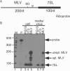
Packaging of 7SL and unspliced, but not spliced, MLV RNAs (RNase protection assay). (A) Riboprobe used. Ψ, packaging signal; SD, splice donor site; thin line, MLV-protected portion of riboprobe; boldface line, 7SL-protected region of riboprobe. (B) Viral and cellular RNAs from uninfected and MLV-infected cells using the 7SL-MLV riboprobe. The positions of the undigested probe, unspliced MLV RNA, spliced MLV RNA, and 7SL RNA bands are indicated on the right (probe, unspl. MLV, spl. MLV, and 7SL, respectively). Lanes: 1, size markers (M); 2, undigested probe; 3, RNA from packaging cells; 4, uninfected cells; 5, infected cells; 6, packaging cell supernatants; 7, uninfected cell supernatants; 8, infected cell supernatants. Bands were quantified by PhosphorImager analysis and normalized for the number of Cs in each protected probe fragment.
FIG. 3.
FIG. 3.
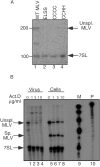
7SL packaging determinants (RNase protection assays). (A) Virion RNAs from wild-type (WT) or NC mutant MLV. Lanes: 1, wild type; 2, ELSS basic region NC mutant (R16L-R17S-R18S); 3, CCCC zinc finger NC mutant (H34C); 4, CCHH zinc finger NC mutant (C39H). 7SL RNA- and MLV genomic RNA-protected product bands are indicated by the arrows. (B) Virion and cellular RNAs from actinomycin D (Act.D)-treated MLV-producing cells. Lanes: 1 to 4, viral RNAs; 5 to 8, cellular RNAs; 9, marker (M); 10, probe (P). Lanes 1 and 5, untreated control; lanes 2 and 6, cells treated with 1 μg/ml actinomycin D; lanes 3 and 7, 5 μg/ml actinomycin D; lanes 4 and 8, 10 μg/ml actinomycin D. 7SL RNA and MLV genomic RNA bands are indicated by the arrows. Note that products migrating between genome and 7SL products in lane 1 are spurious products of RNase digestion.
FIG. 4.
FIG. 4.
Northern blots to assess virion enrichment of ribosomal RNAs and tRNAs. (A) Northern blots using RNA from gradient-purified, micrococcal nuclease-treated MLV probed for 7SL RNA and tRNAs. Left, probed for tRNAPro; middle, probed for tRNAMet; right, probed for tRNAVal. On all blots, lanes loaded right to left are cellular RNA, size markers, and gradient fractions 1 to 23, as indicated below panel B. (B) Northern blots using RNA from gradient-purified, micrococcal nuclease-treated MLV probed for 7SL RNA and rRNAs. Left, probed for 5S rRNA; middle, probed for 5.8S rRNA; right, probed for 18S rRNA. On all blots, lanes are labeled as follows: C, cellular RNA; M, size markers; 1 to 23, gradient fractions. The position of 7SL RNA is indicated by the arrows.
FIG. 5.
FIG. 5.
Northern blots to assess virion enrichment of some other small host RNAs. (A) Northern blots using RNA from gradient-purified, micrococcal nuclease-treated MLV probed for 7SL RNA and cytoplasmic RNAs. Left, probed for Y1 RNA; middle, probed for Y3 RNA; right, probed for B1 RNA. (B) Northern blots using RNA from gradient-purified, micrococcal nuclease-treated MLV probed for 7SL RNA and nuclear RNAs. Left, probed for U6 snRNA; middle, probed for U1 and U5 snRNAs; right, probed for 7SK RNA. On all blots, lanes are labeled as follows: C, cellular RNA; M, size markers; 1 to 23, gradient fractions. The position of 7SL RNA is indicated by the arrows.
FIG. 6.
FIG. 6.
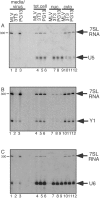
Northern blots of viral, total cell (tot.cell), nuclear (nuc.), and cytoplasmic (cyto) RNAs. Top, probed for 7SL RNA and U5 snRNA; middle, probed for 7SL RNA and Y1 RNA; bottom, probed for 7SL RNA and U6 snRNA. On all blots, lanes 1 to 3 are supernatant/viral RNA, lanes 4 to 6 are total cell RNA, lanes 7 to 9 are nuclear RNA, and lanes 10 to 12 are cytoplasmic RNA.
FIG. 7.
FIG. 7.
Histograms of quantitative RT-PCR data. (A) cellular ratios of unspliced MLV genome cDNA versus actin cDNA and spliced MLV env cDNA; (B) viral ratios of unspliced MLV genome cDNA versus actin cDNA and spliced MLV env cDNA; (C) actin cDNA versus MLV env cDNA. x axis, source of RNA for cDNA synthesis; y axis, ratio of actin to MLV env.

Similar articles

Cited by

References

    1. Adam, M. A., and A. D. Miller. 1988. Identification of a signal in a murine retrovirus that is sufficient for packaging of nonretroviral RNA into virions. J. Virol. 62:3802-3806. - PMC - PubMed
    1. Adkins, B., and T. Hunter. 1981. Identification of packaged cellular mRNA in virions of Rous sarcoma virus. J. Virol. 39:471-480. - PMC - PubMed
    1. Aronoff, R., and M. Linial. 1991. Specificity of retroviral RNA packaging. J. Virol. 65:71-80. - PMC - PubMed
    1. Banks, J. D., B. O. Kealoha, and M. L. Linial. 1999. An MΨ-containing heterologous RNA, but not env mRNA, is efficiently packaged into avian retroviral particles. J. Virol. 73:8926-8933. - PMC - PubMed
    1. Basyuk, E., T. Galli, M. Mougel, J.-M. Blanchard, M. Sitbon, and E. Bertrand. 2003. Retroviral genomic RNAs are transported to the plasma membrane by endosomal vesicles. Dev. Cell 5:161-174. - PubMed

Publication types

MeSH terms