Comparative Study
. 2005 Nov;79(21):13594-605.
doi: 10.1128/JVI.79.21.13594-13605.2005.

Quantitative analysis of the hepatitis C virus replication complex

Affiliations
Comparative Study

Quantitative analysis of the hepatitis C virus replication complex

Doris Quinkert et al. J Virol. 2005 Nov.

Abstract

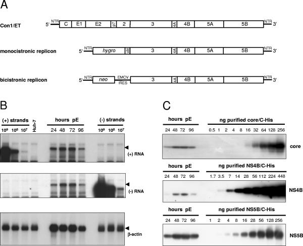
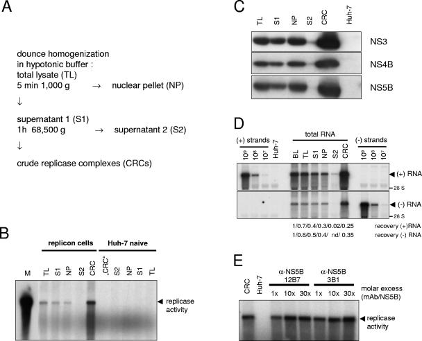
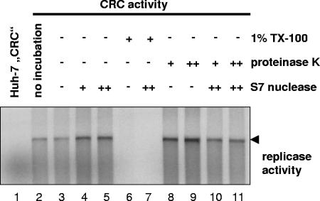
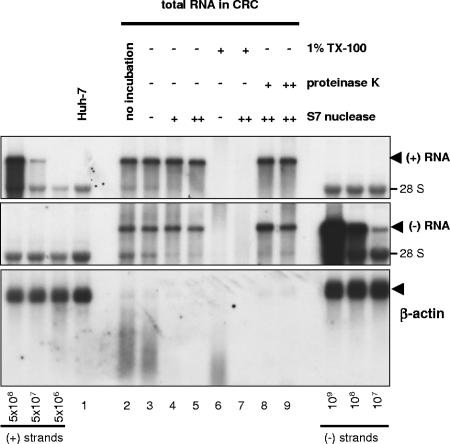
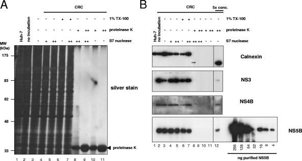
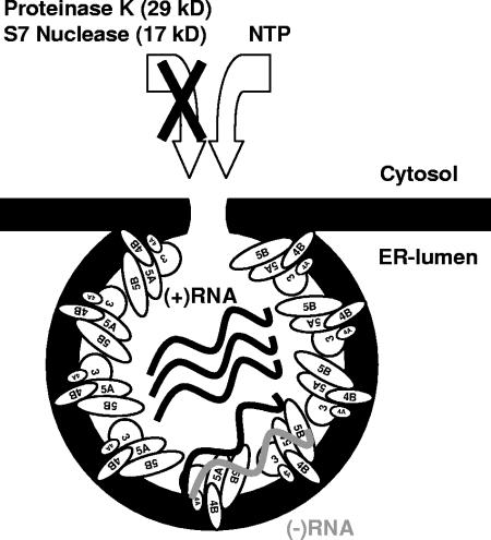
The hepatitis C virus (HCV) encodes a large polyprotein; therefore, all viral proteins are produced in equimolar amounts regardless of their function. The aim of our study was to determine the ratio of nonstructural proteins to RNA that is required for HCV RNA replication. We analyzed Huh-7 cells harboring full-length HCV genomes or subgenomic replicons and found in all cases a >1,000-fold excess of HCV proteins over positive- and negative-strand RNA. To examine whether all nonstructural protein copies are involved in RNA synthesis, we isolated active HCV replication complexes from replicon cells and examined them for their content of viral RNA and proteins before and after treatment with protease and/or nuclease. In vitro replicase activity, as well as almost the entire negative- and positive-strand RNA, was resistant to nuclease treatment, whereas <5% of the nonstructural proteins were protected from protease digest but accounted for the full in vitro replicase activity. In consequence, only a minor fraction of the HCV nonstructural proteins was actively involved in RNA synthesis at a given time point but, due to the high amounts present in replicon cells, still representing a huge excess compared to the viral RNA. Based on the comparison of nuclease-resistant viral RNA to protease-resistant viral proteins, we estimate that an active HCV replicase complex consists of one negative-strand RNA, two to ten positive-strand RNAs, and several hundred nonstructural protein copies, which might be required as structural components of the vesicular compartments that are the site of HCV replication.


Figures

FIG. 1.
FIG. 1.
Quantification of HCV RNA and nonstructural proteins in Huh-7 cells transfected with a full-length genome. (A) Structure of the HCV RNAs used in the present study. Con1/ET represents a full-length HCV genome harboring cell culture-adaptive mutations in NS3 and NS4B (64). The monocistronic replicon contains only a single ORF consisting of nt 342 to 389 of the core-coding region, the hygro gene (encoding the hygromycin phosphotransferase), the ubiquitin-encoding sequence (Ubi), and the HCV NS proteins NS3 to NS5B (24) and was used to select Huh-7 cell clone 11-1, which was analyzed in the present study (Table 1). The bicistronic replicon is composed of the first 377 nt of the HCV genome fused to the neo gene (encoding the neomycin phosphotransferase). Translation of the HCV NS proteins NS3-5B is initiated by the encephalomyocarditis virus-IRES. The bicistronic replicon was used to generate cell-clone 9-13 (53). (B) Time course of HCV positive- and negative-strand synthesis after transfection of Con1/ET RNA into Huh7-Lunet cells. Cells were harvested and counted at the time points indicated above the figure (pE, postelectroporation), and total RNA was prepared. A total of 5 μg of total RNA corresponding to 2.5 × 10⁵, 2.1 × 10⁵, 2.1 × 10⁵, and 2.3 × 10⁵ cells at 24, 48, 72, and 96 h, respectively, was subjected to Northern hybridization with radiolabeled riboprobes specific for the detection of HCV positive-strand (top panel), negative-strand (middle panel) or β-actin (lowest panel). Specific signals are indicated by arrowheads. Signals were quantified by phosphorimaging with known amounts of in vitro transcripts of positive or negative polarity corresponding to a subgenomic replicon and normalized for different loadings by the β-actin signal. Total RNA from naive Huh-7 cells was used as negative control (Huh-7). (C) Quantification of HCV core (upper panel), NS4B (middle panel), and NS5B (lowest panel) expression after transfection of Con1/ET RNA into Huh7-Lunet cells. An aliquot of the cells harvested for total RNA preparation at the time points given above each panel was lysed in Laemmli sample buffer and subjected to immunoblot analysis with monoclonal antibodies (NS5B) or polyclonal antisera (core, NS4B) with the specificities given on the right. Samples were quantified by comparison of signal intensities derived from known amounts of the respective antigens as indicated above each panel. The amount of loaded proteins correspond to 1.2 × 10⁵, 1.8 × 10⁵, 1.6 × 10⁵, and 2.0 × 10⁵ cells at 24, 48, 72, and 96 h, respectively.
FIG. 2.
FIG. 2.
Preparation and characterization of CRCs from HCV replicon cells. (A) Schematic diagram of the CRC preparation protocol. (B) Analysis of in vitro replicase activity in total lysates (TL) and different subcellular fractions of replicon cells (left half) and naive Huh-7 cells (right half). In vitro replicase activity was determined in 4 μl of each fraction, and reaction products were analyzed by denaturing glyoxal-gel electrophoresis, followed by autoradiography of the dried gel. A radioactively labeled in vitro transcript identical in size to the replicon was loaded as a marker (M). The major reaction product of the in vitro replicase assay is indicated by an arrowhead. (C) Detection of NS3, NS4B, and NS5B in different fractions of the CRC preparation. The volume of the NP fraction was adjusted to the volume of S1 and 10 μl of each fraction were analyzed by immunoblot with a polyclonal antiserum raised against HCV NS3 (upper panel) or NS4B (middle panel) or monoclonal antibodies specific for NS5B (lower panel) and compared to 10 μl of “CRC” fraction from naive Huh-7 cells (Huh-7). (D) Fate of viral positive and negative strands during hypotonic lysis and CRC preparation. Total RNA was prepared from 50 μl of a replicon cell suspension before lysis (BL) and from the same volumes of TL, S1, NP (adjusted to the volume of S1), S2, and CRC and subjected to Northern hybridization analysis with the same controls and probes to detect HCV positive- (upper panel) and negative-strand RNA (lower panel) as in Fig. 1B. To calculate the recovery rate samples were analyzed by phosphorimaging and correlated with the value obtained before cell lysis (BL). The data of the CRC fraction were corrected for the difference in total volume. (E) Effect of NS5B-specific monoclonal antibodies on in vitro replicase activity. Two μl of a standard CRC preparation containing 40 ng of NS5B were preincubated 5 min on ice with 0.1, 1, or 3 μg of purified monoclonal antibodies as indicated in the top, resulting in a 1×, 10×, or 30× molar excess or incubated in the absence of antibodies (CRC) and analyzed for in vitro replicase activity. The same amount of “CRC” fraction from naive Huh-7 cells was used as a negative control (Huh-7). For further details refer to the text.
FIG. 3.
FIG. 3.
HCV replicase activity is completely resistant to protease and nuclease treatment. A total of 50 μl of CRCs prepared from replicon cell clone 9-13 was incubated for 60 min at 25°C in the presence of 1% Triton X-100 and/or 0.8 (+) or 8 (++) mg of proteinase K/ml and/or 0.2 (+) or 2 (++) U of S7 nuclease/μl, respectively, as indicated above each lane. After termination of the proteinase K and S7 nuclease digest by the addition of 1.4 mM PMSF and/or 2.75 mM EGTA, respectively, equal amounts of each sample were analyzed for in vitro replicase activity. Reaction products were separated by denaturing glyoxal agarose gel electrophoresis and autoradiography. Lane 1 and 2 represent control reactions with CRCs from naive Huh-7 or replicon cells in the absence of any preincubation. CRCs in lane 3 were mock incubated for 60 min at 25°C prior to the in vitro replicase assay.
FIG. 4.
FIG. 4.
Quantification of nuclease-resistant HCV positive- and negative-strand RNA and β-actin mRNA in CRCs. Total RNA equivalent to 5 μl of CRCs treated with 1% Triton X-100, 0.8 (+) or 8 (++) mg of proteinase K/ml and/or 0.2 (+) or 2 (++) U of S7 nuclease/μl for 60 min at 25°C or from mock-treated CRCs, as indicated on top, was subjected to Northern hybridization analysis with a negative-strand riboprobe to detect viral positive-strand RNA (top panel), a positive-strand riboprobe for HCV negative-strand detection (middle panel), and a riboprobe specific for the detection of cellular β-actin mRNA. The positions of viral positive- and negative-strand RNA and β-actin are indicated by arrowheads at the right. For quantification, in vitro transcribed replicons corresponding to known amounts of viral positive- and negative-strand RNA were mixed with 2 μg of total cellular RNA from naive Huh-7 cells and loaded as indicated at the bottom of the figure. We used 4 μg of total RNA from naive Huh-7 cells as a negative control (Huh-7, lane 1). We quantified the viral RNAs by phosphorimaging and found ca. 3 × 10⁶ negative-strand and 3 × 10⁷ positive-strand RNAs per μl of CRCs in this particular experiment.
FIG. 5.
FIG. 5.
Effect of proteinase K digest on cellular proteins and quantification of protease-resistant HCV nonstructural proteins in CRCs. Equal amounts of CRCs prepared from replicon cell clone 9-13 were incubated for 60 min at 25°C in the presence or absence of 1% Triton X-100 and/or 0.8 (+) or 8 (++) mg of proteinase K/ml and/or 0.2 (+) or 2 (++) U of S7 nuclease/μl, respectively, as indicated above each lane. The reaction was stopped by the addition of 1.4 mM PMSF and 2.75 mM EGTA and boiled in sample buffer, and total protein equivalent to 2 μl of the CRCs was subjected to SDS-10% PAGE. In the fivefold-concentrated samples, the proteins were trichloroacetic acid precipitated, and the equivalent of 10 μl of CRCs was loaded. Proteins were either visualized by silver staining (A) or subjected to immunoblot analysis (B) with monoclonal antibodies specific for the ER luminal part of calnexin (upper panel), a polyclonal antiserum raised against HCV NS3 or NS4B (middle two panels), and monoclonal antibodies specific for HCV NS5B (bottom panel), as indicated at the right. The concentrated fractions (lane 12) are shown from identical expositions of the same blot. A serial dilution of purified NS5B was loaded in parallel for quantification as depicted at the bottom of the figure.
FIG. 6.
FIG. 6.
Schematic model of the HCV replication complex. HCV NS proteins are indicated by ellipses; black and gray wavy lines represent viral positive- and negative-strand RNAs, respectively. Individual NS proteins and RNA are not drawn to scale. For closer explanations refer to the text.

References

    1. Adachi, T., H. Ago, N. Habuka, K. Okuda, M. Komatsu, S. Ikeda, and K. Yatsunami. 2002. The essential role of C-terminal residues in regulating the activity of hepatitis C virus RNA-dependent RNA polymerase. Biochim. Biophys. Acta 1601:38-48. - PubMed
    1. Aizaki, H., K. J. Lee, V. M. Sung, H. Ishiko, and M. M. Lai. 2004. Characterization of the hepatitis C virus RNA replication complex associated with lipid rafts. Virology 324:450-461. - PubMed
    1. Ali, N., and A. Siddiqui. 1995. Interaction of polypyrimidine tract-binding protein with the 5′ noncoding region of the hepatitis C virus RNA genome and its functional requirement in internal initiation of translation. J. Virol. 69:6367-6375. - PMC - PubMed
    1. Ali, N., and A. Siddiqui. 1997. The La antigen binds 5′ noncoding region of the hepatitis C virus RNA in the context of the initiator AUG codon and stimulates internal ribosome entry site-mediated translation. Proc. Natl. Acad. Sci. USA 94:2249-2254. - PMC - PubMed
    1. Ali, N., K. D. Tardif, and A. Siddiqui. 2002. Cell-free replication of the hepatitis C virus subgenomic replicon. J. Virol. 76:12001-12007. - PMC - PubMed

Publication types

LinkOut - more resources