. 2005 Oct 25;102(43):15494-9.
doi: 10.1073/pnas.0507612102. Epub 2005 Oct 14.

Functions and dysfunctions of the nuclear lamin Ig-fold domain in nuclear assembly, growth, and Emery-Dreifuss muscular dystrophy

Affiliations

Functions and dysfunctions of the nuclear lamin Ig-fold domain in nuclear assembly, growth, and Emery-Dreifuss muscular dystrophy

Dale K Shumaker et al. Proc Natl Acad Sci U S A. .

Abstract

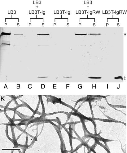
The non-alpha-helical C terminus of Xenopus lamin B3 (LB3T) inhibits the polymerization of lamin B3 in vitro and prevents the assembly of nuclei in Xenopus egg interphase extracts. To more precisely define the functions of LB3T in nuclear assembly, we have expressed subdomains of LB3T and determined their effects on nuclear assembly in Xenopus extracts. The results demonstrate that the Ig-fold motif (LB3T-Ig) is sufficient to inhibit lamin polymerization in vitro. Addition of the LB3T-Ig to egg extracts before the introduction of chromatin prevents chromatin decondensation and the assembly of the lamina, membranes, and pore complexes comprising the nuclear envelope. When added to assembled nuclei, LB3T-Ig prevents the further incorporation of lamin B3 into the endogenous lamina and blocks nuclear growth. The introduction of a point mutation in LB3T-Ig (R454W; LB3T-IgRW), known to cause Emery-Dreifuss muscular dystrophy when present in lamin A, does not inhibit lamin polymerization, chromatin decondensation, or nuclear assembly and growth. These results shed light on the specific alterations in lamin functions attributable to a known muscular dystrophy mutation and provide an experimental framework for revealing the effects of other mutations causing a wide range of laminopathies.


Figures

Fig. 1.
Fig. 1.
The Ig-fold subdomain is sufficient to inhibit nuclear assembly. The structure of lamin B3 is shown with red boxes for the coil-coiled central rod domain and blue arrows indicating the β-strands comprising the Ig-fold. Also shown is an expanded view of the C-terminal tail domain with some amino acid positions indicated. The numbers on the bars of the graph refer to the amino acid residues of lamin B3 fragments added to interphase Xenopus extracts. The nuclear area was determined from the midsection of nuclei by using Zeiss lsm software (n = 100), as described in Materials and Methods. The gray bars indicate the subdomains that inhibited nuclear assembly.
Fig. 2.
Fig. 2.
The Ig-fold inhibits nuclear membrane and lamina assembly and chromatin decondensation. Sperm chromatin was incubated in extracts containing LB3T-Ig (AC), LB3T-58A (DF), LB3T-58B (GI), LB3T-52 (JL), lamin B3 (MO), and LB3-IgRW (PR) for 90 min (see Materials and Methods). The resulting preparations were stained with the DNA dye TOTO (A, D, G, J, M, and P), the lipophilic dye DiOC6 (B, E, H, K, N, and Q), and a mAb directed against lamin B3 (C, F, I, L, O, and R). Chromatin in extracts containing LB3T-Ig (10 μM) remained highly condensed (A), surrounded by small patches of lamins and membranes (B and C). However, chromatin incubated in extracts containing LB3T-58A, LB3T-58B, LB3T-52, lamin B3, or LB3T-IgRW (all 10 μM) were decondensed (D, G, J, M, and P) and surrounded by rims of lamin and membrane fluorescence (E, F, H, I, K, L, N, O, Q, and R). All images were captured from confocal sections taken through the midregions of nuclei. (Scale bar, 10 μm.)
Fig. 3.
Fig. 3.
The Ig-fold inhibits lamin polymerization in vitro. Purified lamin B3 (A and B), LB3T-Ig (E and F), or LB3T-IgRW (I and J) were diluted to a final concentration of 0.6 μM in LAB at 22°C. After 30 min, each of these preparations were centrifuged at 20,000 × g for 20 min. The supernatants and pellets were analyzed by SDS/PAGE. Under these conditions, ≈95% of the lamin B3 was detected in the pellet (A and B), whereas LB3T-Ig (E and F) and LB3T-IgRW (I and J) remained in the supernatant. When lamin B3 was mixed with LB3T-Ig (1:3 molar ratio; C and D), however, the majority of lamin B3 remained in the supernatant (C and D). In the presence of LB3T-IgRW (1:5 molar ratio), the majority of lamin B3 was found in the pellet fraction (G and H). Purified lamin B3 at a concentration of 0.6 μMin LAB was negatively stained with 1% uranyl acetate (K). The spacing of the paracrystal repeats is ≈24 nm. *, lamin B3; ‡, Ig-fold protein. (Scale bar, 250 nm.)
Fig. 4.
Fig. 4.
Nuclei were assembled in Xenopus interphase extract for 45 min. The resulting nuclei were treated with 0.1% Nonidet P-40 in NWB containing lamin B3, His-LB3T-Ig, or His-LB3T-IgRW for 10 min (see Materials and Methods). The nuclei were then washed in NWB, centrifuged, suspended in fresh interphase extract to reseal their membranes, and grown for an additional 60 min (see Materials and Methods). Under these conditions, the average increase in nuclear area was >90% relative to their size at 45 min after nuclear assembly was initiated. Nuclei exposed to LB3T increased in size by only ≈12%, whereas nuclei treated with LB3T-Ig grew ≈2%. In contrast, the LB3T-IgRW-treated nuclei grew ≈82% (n = 50).
Fig. 5.
Fig. 5.
Nuclei were assembled for 45 min, permeabilized with 0.1% Nonidet P-40 in the presence of either His-LB3T-Ig or His-LB3T-IgRW, and transferred to fresh interphase extract for 60 min (see Materials and Methods). Nuclei loaded with His-LB3T-Ig and stained with a mAb directed against His indicate that LB3T-Ig is incorporated into the nuclear lamina and nucleoplasm (A). In some cases, permeabilized nuclei loaded with either LB3T-Ig or LB3T-IgRW were resuspended in fresh interphase extract containing FLAG-LB3 and grown for an additional 60 min. Immunoblot analyses of these nuclei showed that LB3T-Ig significantly reduced the incorporation of FLAG-LB3 when compared with nuclei loaded with LB3T-RW (B). Preparations of these nuclei were also fixed and stained with TOTO (C and F), a mAb directed against lamin B3 (D and G), and a pAb directed against the FLAG epitope (E and H). Lamin B3 was present in nuclei treated with LB3T-Ig (D), but these nuclei did not incorporate FLAG-LB3 (E) into the lamina, whereas nuclei treated with LB3T-IgRW incorporated FLAG-LB3 (H) into the lamina. (Scale bars, 10 μm.)

References

    1. Riemer, D., Wang, J., Zimek, A., Swalla, B. J. & Weber, K. (2000) Gene 255, 317-325. - PubMed
    1. Karabinos, A., Zimek, A. & Weber, K. (2004) Gene 326, 123-129. - PubMed
    1. Gruenbaum, Y., Margalit, A., Goldman, R. D., Shumaker, D. K. & Wilson, K. L. (2005) Nat. Rev. Mol. Cell Biol. 6, 21-31. - PubMed
    1. Hesse, M., Magin, T. M. & Weber, K. (2001) J. Cell Sci. 114, 2569-2575. - PubMed
    1. Strelkov, S. V., Schumacher, J., Burkhard, P., Aebi, U. & Herrmann, H. (2004) J. Mol. Biol. 343, 1067-1080. - PubMed

Publication types