. 2005 Nov;25(21):9198-208.
doi: 10.1128/MCB.25.21.9198-9208.2005.

Micro-RNA regulation of the mammalian lin-28 gene during neuronal differentiation of embryonal carcinoma cells

Affiliations

Micro-RNA regulation of the mammalian lin-28 gene during neuronal differentiation of embryonal carcinoma cells

Ligang Wu et al. Mol Cell Biol. 2005 Nov.

Abstract

Vertebrate genomes each encode hundreds of micro-RNAs (miRNAs), yet for few of these miRNAs is there empirical evidence as to which mRNA(s) they regulate. Here we report the identification of human lin-28 mRNA as a regulatory target of human miR-125b and its homolog miR-125a. Studies of miR-125b function in mouse P19 embryonal carcinoma cells induced to develop into neurons suggest a role for this regulatory miRNA in mammalian neuronal differentiation, since its increased concentration in these cells contributes to lin-28 downregulation. Within the lin-28 3' untranslated region (UTR) are two conserved miRNA responsive elements (miREs) that mediate repression by miR-125b and miR-125a. Simultaneous deletion of both miREs renders the lin-28 3' UTR almost completely insensitive to these miRNAs, indicating that these two miREs are the principal elements in the lin-28 3' UTR that respond to miR-125. At the 3' end of each element is an adenosine residue that makes a significant contribution to function irrespective of its complementarity to the 5'-terminal nucleotide of miR-125. By contrast to most earlier reports of gene repression by other miRNAs that are imperfectly complementary to their targets, lin-28 downregulation by miR-125 involves reductions in both translational efficiency and mRNA abundance. The decrease in the mRNA concentration is achieved by a posttranscriptional mechanism that is independent of the inhibitory effect on translation.


Figures

FIG. 1.
FIG. 1.
Human miR-125a and miR-125b. A. Sequence of miR-125a, miR-125b, and C. elegans lin-4 miRNA. The miRNAs are drawn in a 5′-to-3′ orientation (left to right), and the conserved regions are highlighted. B. Increased abundance of miR-125a and miR-125b in transfected cells. 293T cells were transiently transfected either with a plasmid (pMIR125a or pMIR125b) encoding miR-125a or miR-125b or with a plasmid deletion variant (pMIR125aΔ or pMIR125bΔ) that did not encode an miRNA. After 36 h, total RNA was extracted and analyzed by gel electrophoresis and blotting, using radiolabeled oligonucleotide probes complementary to miR-125a or miR-125b. The sequence similarity of miR-125a and miR-125b resulted in some cross-hybridization of the two probes. miR-125a migrated as a doublet, suggesting two forms differing in length by one nucleotide, perhaps indicative of heterogeneity at the 3′-terminal processing site (±A). The sizes of the larger RNAs that were also detected suggest that they are partially processed stem-loop precursors of miR-125a and miR-125b (pre-miR-125a and pre-miR-125b). Unlike the miRNAs and pre-miRNAs in the cell extracts, the oligoribonucleotides used as approximate size markers (M) were not phosphorylated at the 5′ end and therefore migrated somewhat more slowly during electrophoresis than they would have otherwise.
FIG. 2.
FIG. 2.
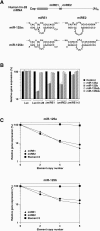
Repression of luciferase reporters bearing lin-28 miREs by miR-125a and miR-125b. A. Map of human lin-28 mRNA and duplexes of the lin-28 miREs with miR-125a and miR-125b. In the RNA duplexes, the miRE (top strand) is drawn in a 5′-to-3′ orientation (left to right), and the miRNA (bottom strand) is drawn in a 3′-to-5′ orientation (left to right). These two lin-28 miREs are located directly beside one another, such that the two nucleotides shown at the 3′ end of miRE1 and at the 5′ end of miRE2 are the same two nucleotides of the lin-28 3′ UTR. B. Repression of a luciferase reporter bearing the lin-28 3′ UTR by miR-125a and miR-125b. 293T cells were transiently cotransfected with a luciferase reporter plasmid bearing either the human lin-28 3′ UTR (Luc-lin 28) or the SV40 late 3′ UTR (Luc), together with a plasmid encoding β-galactosidase (an internal standard to control for transfection efficiency) and one of five plasmids encoding either miR-125a or miR-125b, deletion variants thereof, or no miRNA-related transcript (control). Alternatively, the cotransfections were performed using Luc-lin 28 reporters from which miRE1 and/or miRE2 had been deleted. After 36 h, the ratio of luciferase activity to β-galactosidase activity in cell extracts was measured. Error bars correspond to the standard deviation of multiple measurements. C. Repression of a luciferase reporter bearing multiple copies of the lin-28 miREs by miR-125a and miR-125b. 293T cells were transiently cotransfected with a plasmid containing a luciferase-SV40 reporter gene (Luc) that bore 0, 2, 4, or 6 copies of miRE1, miRE2, or lin-28 element X (UGCAAGUGAGGGUUCUGGGGG) in its 3′ UTR and with one of four plasmids encoding miR-125a or miR-125b or deletion variants thereof, together with a plasmid encoding β-galactosidase (internal standard). After 36 h, the ratio of luciferase activity to β-galactosidase activity in cell extracts was measured. The activity ratio for cells transfected with genes encoding miR-125a or miR-125b was then normalized to the ratio in cells transfected with the corresponding deletion mutant that did not encode an miRNA and graphed.
FIG. 3.
FIG. 3.
Nonresponsive or poorly responsive elements in the lin-28 3′ UTR. Hypothetical duplexes of nonresponsive or poorly responsive lin-28 3′ UTR elements (top strands, 5′-to-3′ orientation) with miR-125b (bottom strands, 3′-to-5′ orientation). The relative potency of each element in mediating translational repression by miR-125b in transfected 293T cells was calculated as (RE − 1)/(RmiRE1 − 1), where R is the ratio of luciferase production (normalized to β-galactosidase activity) in the absence versus the presence of miR-125b for a reporter bearing either two copies of the element (RE) or two copies of miRE1 (RmiRE1). Of the three elements shown, only miRE0 has detectable repression activity, and its effect is only 30% that of miRE1.
FIG. 4.
FIG. 4.
Efficacy of miRE1 variants in repressing gene expression. A. Duplexes of miRE1 and variants thereof (upper strands) with miR-125b (lower strands). Nucleotides in both strands of these duplexes are numbered from right to left. The miRE nucleotides expected to form consecutive base pairs with the 5′ portion of miR-125b are indicated beside each duplex, along with the identity of the nucleotide at the miRE 3′ end (position 1). The miRNA variant miR-125b-U1A (not shown) is identical to miR-125b except for a U→A substitution at the 5′ terminus (position 1). B. Repression of a luciferase-SV40 reporter gene bearing two copies of miRE1 or variants thereof. 293T cells were transiently cotransfected with plasmids encoding a reporter mRNA, β-galactosidase (internal standard), and either miR-125b, an miR-125b substitution mutant bearing a 5′terminal adenosine (miR-125b-U1A), or an miR-125b deletion mutant (miR-125bΔ). After 36 h, the ratio of luciferase activity to β-galactosidase activity in cell extracts was measured. By dividing the ratio in cells lacking miR-125b by the ratio in otherwise identical cells containing miR-125b (black bars) or miR-125b-U1A (gray bars), repression ratios were calculated for each 3′ UTR element. These repression ratios were then graphed on a logarithmic scale.
FIG. 5.
FIG. 5.
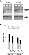
Relative contributions of translational efficiency and mRNA abundance to repression by miR-125a and miR-125b. A. RNA blot showing the effect of miR-125a and miR-125b on the cellular concentration of a reporter mRNA containing zero or six copies of lin-28 miRE1 or miRE2. 293T cells were transiently cotransfected with a reporter gene, a β-galactosidase gene, and a gene encoding miR-125a (a), miR-125b (b), or an otherwise identical primary transcript from which the pre-miRNA stem-loop had been deleted (Δ). After 36 h, cytoplasmic RNA was isolated and analyzed by gel electrophoresis and blotting, using radiolabeled probes complementary to luciferase mRNA and β-galactosidase mRNA (internal standard). B. Contributions of translational inhibition and diminished mRNA abundance to repression by miR-125a and miR-125b. The reporter mRNA concentration in each sample was normalized to the concentration of β-galactosidase mRNA, and the ratio of the normalized concentration of the reporter mRNA in cells transfected with an miR-125a or miR-125b gene versus the corresponding miRNA deletion variant was calculated. To reveal the relative contributions of translational efficiency (protein yield per mRNA molecule) and mRNA abundance to repressing gene expression, the calculated effect of each miRNA on the cytoplasmic abundance of reporter mRNAs bearing six copies of miRE1 or miRE2 was superposed on a graph showing the overall degree of repression of the same reporter genes by each miRNA. This overall degree of repression, the repression ratio, was calculated as for Fig. 4. The effect of an miRNA on translational efficiency (black bars) corresponds to the ratio of its overall effect on protein synthesis versus its effect on mRNA abundance (gray bars). In the case of miRE1, expression of miR-125a or miR-125b reduced translational efficiency by a factor of 4.3 ± 0.3 or 2.9 ± 0.4, respectively, and mRNA abundance by a factor of 4.2 ± 0.2 or 3.2 ± 0.1, respectively. In the case of miRE2, expression of miR-125a or miR-125b reduced translational efficiency by a factor of 3.8 ± 0.4 or 2.2 ± 0.3, respectively, and mRNA abundance by a factor of 3.1 ± 0.3 or 2.7 ± 0.3, respectively.
FIG. 6.
FIG. 6.
Consequences of inserting a polyadenylation site upstream of the miREs. A. Luciferase reporter gene bearing an intact (wt: AATAAA) or defective (mut: TTCTTT) bovine growth hormone (BGH) polyadenylation signal upstream of six copies of lin-28 miRE1 and an intact SV40 polyadenylation signal downstream of the miREs. B. Effect of miR-125b on the expression of reporter genes in which an intact (wt) or defective (mut) BGH polyadenylation signal had been inserted into the 3′ UTR upstream of zero or six copies of lin-28 miRE1. 293T cells were transiently cotransfected with a reporter gene, a β-galactosidase gene, and a gene encoding miR-125b or an otherwise identical primary transcript from which the pre-miRNA stem-loop had been deleted (miR-125bΔ). After 36 h, the ratio of luciferase activity to β-galactosidase activity in cell extracts was measured. C. RNA blot showing the effect of miR-125b on the cytoplasmic concentration of a reporter mRNA in which an intact (wt) or defective (mut) BGH polyadenylation signal had been inserted upstream of six copies of lin-28 miRE1. 293T cells were transiently cotransfected as in A, and after 36 h, cytoplasmic RNA was isolated and analyzed as for Fig. 5. Compared to a negative control (Δ = miR-125bΔ), miR-125b (b) reduced the cytoplasmic concentration of the 2.4-kb reporter mRNA bearing a defective polyadenylation signal and six copies of miRE1 to 44% ± 4% of its unrepressed value but did not affect the concentration of the 1.8-kb mRNA transcribed from an otherwise identical gene bearing a functional polyadenylation signal upstream of the miREs (98% ± 7%).
FIG. 7.
FIG. 7.
Translation independence of the downregulation of mRNA abundance by miR-125b. 293T cells were transiently cotransfected with a luciferase reporter gene bearing six copies of miRE1 in the 3′ UTR, with or without a segment encoding a large stem-loop structure (CGGGGCGCGUGGUGGCGGCUGCAGCCGCCACCACGCGCCCCG) in the 5′ UTR, a β-galactosidase gene, and a gene encoding miR-125b (b) or an otherwise identical primary transcript from which the pre-miRNA stem-loop had been deleted (Δ). After 36 h, cytoplasmic RNA was isolated and analyzed as for Fig. 5. The ratio of the reporter mRNA to β-galactosidase mRNA (internal standard) is indicated at the bottom of each lane.
FIG. 8.
FIG. 8.
Changes in miR-125a and miR-125b levels and lin-28 gene expression following retinoic acid induction of P19 cell differentiation. P19 cells were grown for 5 days in agarose-coated petri dishes in the presence or absence of retinoic acid (1 μM). RNA and protein samples were extracted daily, and equal amounts were analyzed by blotting, using radiolabeled DNA probes to detect specific RNAs (miR-125a, miR-125b, and lin-28 mRNA) and polyclonal anti-Lin-28 antibodies to detect Lin-28. tRNA, 18S rRNA, and actin served as internal standards. Lane M, RNA extracted from 293T cells transiently transfected with either pMIR125a (top pair of panels; detection with an miR-125a-specific probe) or pMIR125b (second pair of panels; detection with an miR-125b-specific probe); note the similar abundance of miR-125b in P19 cells induced for 5 days and in transfected 293T cells. Due to low-level cross-hybridization, miR-125b (faint lower band) was detectable with the miR-125a-specific probe, and miR-125a (faint upper band) was detectable with the miR-125b-specific probe.
FIG. 9.
FIG. 9.
Downregulation of lin-28 gene expression by production of miR-125b in undifferentiated P19 cells. Undifferentiated P19 cells were transfected with a plasmid (pSH-MIR125b, 1.5 μg) encoding miR-125b or with a variant thereof (pSHAG-1, 1.5 μg) lacking the miR-125b gene segment (Δ), and after two days, RNA and protein extracts were prepared. Alternatively, untransfected P19 cells (−) were treated with retinoic acid for 4 days, and RNA was extracted 1 day later. Equal amounts of each RNA or protein sample were analyzed by blotting to detect miR-125b, Lin-28 protein, and lin-28 mRNA. tRNA, 18S rRNA, and actin served as internal standards. The increased production of miR-125b in undifferentiated P19 cells that had been transfected with pSH-MIR125b reduced the abundance of the Lin-28 protein and lin-28 mRNA to approximately one-quarter (26% ± 2%) and one-half (52% ± 2%) of their respective concentrations in control cells (Δ).
FIG. 10.
FIG. 10.
Influence of miR-125b on Lin-28 protein synthesis in differentiating P19 cells. A. (Top) Sequence of miR-125b and of the complementary 2′-O-methylated oligonucleotide (2′ OMe anti-miR-125b) used to repress its activity. Also shown is the sequence of a noncomplementary 2′-O-methylated oligonucleotide used as a negative control. (Bottom) The efficacy of the 2′ OMe anti-miR-125b oligonucleotide in repressing miR-125b function was confirmed by transiently cotransfecting undifferentiated P19 cells with a luciferase-SV40 reporter gene bearing zero or six copies of miRE2 in its 3′ UTR (25 ng), a plasmid encoding miR-125b or a deletion variant thereof (750 ng), a plasmid encoding β-galactosidase (25 ng, internal standard), and either the 2′ OMe anti-miR-125b oligonucleotide or the control oligonucleotide (100 pmol). After 48 h, cell extracts were prepared, and the ratio of luciferase activity to β-galactosidase activity was measured. The activity ratio in cells transfected with a reporter containing six copies of miRE2 was then normalized to the ratio in cells transfected with the corresponding reporter lacking this element, and the ratios were graphed. B. Undifferentiated P19 cells were transiently transfected with the 2′ OMe anti-miR-125b oligonucleotide or the negative control 2′ OMe oligonucleotide (200 pmol) and then induced to differentiate by treatment with retinoic acid. Five-and-a-half days after induction, protein extracts were prepared, and equal amounts of each extract were analyzed by immunoblotting to detect Lin-28 protein and actin (an internal standard). Compared to the control oligonucleotide, transfection with the 2′-O-methylated anti-miR-125b oligonucleotide increased Lin-28 protein synthesis by almost a factor of 2 (1.8 ± 0.3).
FIG. 11.
FIG. 11.
Contribution of lin-28 miREs to repressing gene expression in differentiating P19 cells. A. P19 cells that had (+) or had not (−) been treated with retinoic acid were transiently cotransfected with a plasmid encoding a luciferase reporter fused to the human lin-28 3′ UTR (Luc-lin 28, 0.05 μg), a plasmid encoding β-galactosidase (0.05 μg, an internal standard), and pUC19 (0.9 μg). Alternatively, the cotransfection was performed using Luc-lin 28 reporters from which elements responsive to miR-125 (miRE0, miRE1, and miRE2) and/or let-7 miRNA (L7: GAGUGCACAGCCUAUUGAACUACCUCA) had been deleted (see map of human lin-28 mRNA above graph). After 36 h, cell extracts were prepared, and the ratio of luciferase activity to β-galactosidase activity was measured. Prior to graphing, the ratios were normalized to the values measured for the reporter lacking all four miREs. B. P19 cells were transiently cotransfected with a plasmid encoding a luciferase-SV40 reporter mRNA that bore zero or six copies of miRE1 in its 3′ UTR (0.7 μg) and a plasmid encoding β-galactosidase (0.9 μg, internal standard). After 12 h, the cells were transferred to petri dishes and cultured for 4 days in α-MEM containing fetal bovine serum (FBS) (10%) and retinoic acid (1 μM). The cells were then transferred to tissue culture dishes coated with poly-d-lysine and cultured for six more days in either α-MEM containing 10% fetal bovine serum (to allow the growth of nonneuronal cells) or Neurobasal medium containing B27 supplement (to suppress the growth of nonneuronal cells) (4). Cell extracts were prepared at 2-day intervals, and the ratio of luciferase activity to β-galactosidase activity was measured. The activity ratio in cells transfected with the reporter that contained six copies of miRE1 was then normalized to the ratio in cells transfected with the reporter that lacked such miREs, and the data were graphed.

Similar articles

Cited by

References

    1. Ambros, V., and H. R. Horvitz. 1984. Heterochronic mutants of the nematode Caenorhabditis elegans. Science 226:409-416. - PubMed
    1. Berezikov, E., V. Guryev, J. van de Belt, E. Wienholds, R. H. Plasterk, and E. Cuppen. 2005. Phylogenetic shadowing and computational identification of human microRNA genes. Cell 120:21-24. - PubMed
    1. Brennecke, J., A. Stark, R. B. Russell, and S. M. Cohen. 2005. Principles of microRNA-target recognition. PloS. Biology 3:e85. - PMC - PubMed
    1. Brewer, G. J., J. R. Torricelli, E. K. Evege, and P. J. Price. 1993. Optimized survival of hippocampal neurons in B27-supplemented Neurobasal, a new serum-free medium combination. J. Neurosci. Res. 35:567-576. - PubMed
    1. Doench, J. G., C. P. Petersen, and P. A. Sharp. 2003. siRNAs can function as miRNAs. Genes Dev. 17:438-442. - PMC - PubMed

Publication types

LinkOut - more resources