. 2005 Nov;25(21):9304-17.
doi: 10.1128/MCB.25.21.9304-9317.2005.

Transforming growth factor beta2 is a neuronal death-inducing ligand for amyloid-beta precursor protein

Affiliations

Transforming growth factor beta2 is a neuronal death-inducing ligand for amyloid-beta precursor protein

Yuichi Hashimoto et al. Mol Cell Biol. 2005 Nov.

Abstract

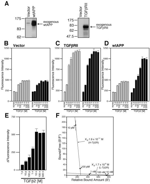
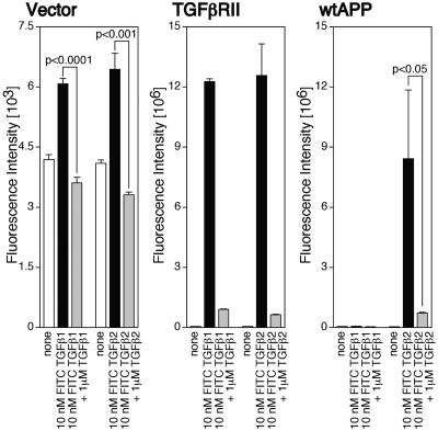
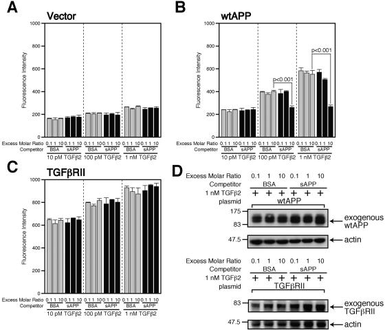
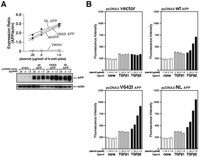
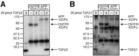
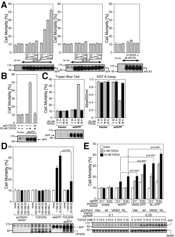
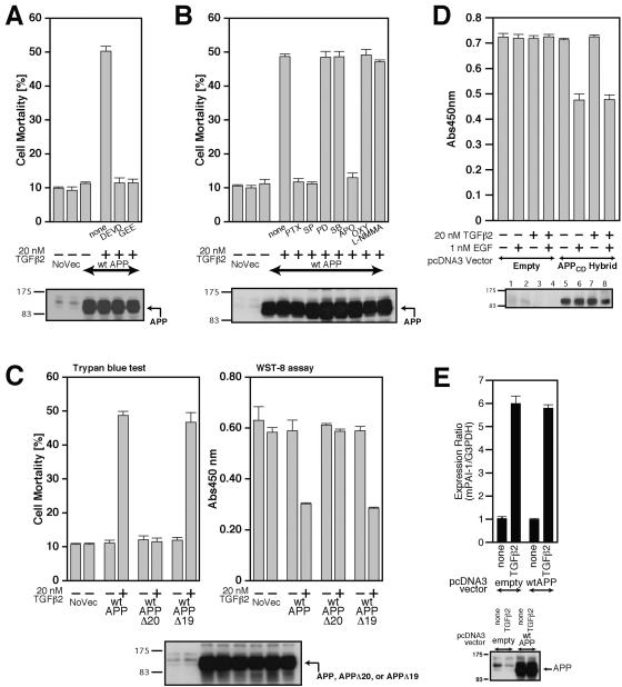
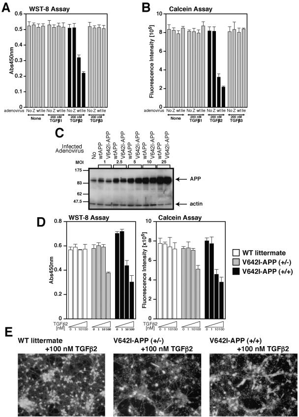
APP, amyloid beta precursor protein, is linked to the onset of Alzheimer's disease (AD). We have here found that transforming growth factor beta2 (TGFbeta2), but not TGFbeta1, binds to APP. The binding affinity of TGFbeta2 to APP is lower than the binding affinity of TGFbeta2 to the TGFbeta receptor. On binding to APP, TGFbeta2 activates an APP-mediated death pathway via heterotrimeric G protein G(o), c-Jun N-terminal kinase, NADPH oxidase, and caspase 3 and/or related caspases. Overall degrees of TGFbeta2-induced death are larger in cells expressing a familial AD-related mutant APP than in those expressing wild-type APP. Consequently, superphysiological concentrations of TGFbeta2 induce neuronal death in primary cortical neurons, whose one allele of the APP gene is knocked in with the V642I mutation. Combined with the finding indicated by several earlier reports that both neural and glial expression of TGFbeta2 was upregulated in AD brains, it is speculated that TGFbeta2 may contribute to the development of AD-related neuronal cell death.


Figures

FIG. 1.
FIG. 1.
Overexpression of wtAPP induces the expression of TGFβ2-specific receptors. (A) F11 cells (7 × 10⁴ cells/well in 6-well plates) were transfected with 0.5 μg of the pcDNA3 vector, pcDNA3-TGFβRII, or pcDNA3-wtAPP. Cell lysates (10 μg in each lane) were immunoblotted with antibody to APP (22C11) or TGFβRII. (B to D) F11 cells (7 × 10⁴ cells/well in 6-well plates) were transfected with 0.25 μg of the pcDNA3 vector (B), pcDNA3-TGFβRII (C), or pcDNA3-wtAPP (D). At 36 h after transfection, the cells were combined with the indicated concentrations of TGFβ1 or TGFβ2. Immunofluorescence-based binding assays were performed as described in Materials and Methods. (E) The mean of immunofluorescence intensity numbers representing the association between TGFβ2 and F11 cells for each concentration of TGFβ2 shown in panel B were subtracted from immunofluorescence intensity numbers representing the association between TGFβ2 and F11 cells for the same concentration of TGFβ2 shown in panel D. Resulting intensity numbers were considered to correspond to the association between TGFβ2 and wtAPP-induced TGFβ2-specific receptors. (F) A simulated Scatchard plot of the association between TGFβ2 and wtAPP-induced TGFβ2-specific receptors based on the analysis shown in Table 1. A vertical bar for each point indicates an estimated range of bound/free numbers. p, pico; n, nano; B′, relative amount of bound TGFβ2; F′, estimated amount of free TGFβ2.
FIG. 2.
FIG. 2.
Specific association between TGFβ2 and F11 cells overexpressing wtAPP. F11 cells (7 × 10⁴ cells/well in 6-well plates) were transfected with 0.25 μg of the TGFβRII-encoding plasmid or the wtAPP-encoding plasmid and were replated into 96-well plates coated with poly-l-lysine (7 × 10³ cells/well) at 24 h after transfection. At 36 h after transfection, cells were combined with 10 nM FITC-labeled TGFβ1 or TGFβ2 in the presence or the absence of 1 μM unlabeled TGFβ1 or TGFβ2. To keep the total FITC amounts constant, proper amounts of free FITC were added. After incubation for 6 h, they were washed with PBS three times. The immunofluorescence intensity was measured (excitation, 485 nm; emission, 535 nm) with a spectrofluorometer (Wallac 1420 ARVOsx multilabel counter).
FIG. 3.
FIG. 3.
Soluble APP antagonized TGFβ2-induced death. (A to C) F11 cells (7 × 10⁴ cells/well in 6-well plates) were transfected with 0.25 μg of the pcDNA3 vector (A), pcDNA3-wtAPP (B), or pcDNA3-TGFβRII (C). At 36 h after transfection, cells were combined with the indicated concentrations of TGFβ1 or TGFβ2 in the presence of the indicated amounts of sAPP or BSA as a competitor. Immunofluorescence-based binding assays were performed as described in Materials and Methods. (D) F11 cells (7 × 10⁴ cells/well in 6-well plates) were transfected with 0.25 μg of pcDNA3-wtAPP (upper panel) or pcDNA3-TGFβRII (lower panel). At 36 h after transfection, cells were combined with 1 nM TGFβ2 in the presence of 10 nM sAPP or BSA. Cell lysates (10 μg in each lane) were immunoblotted with antibody to APP (22C11) (upper) or TGFβRII (lower) as well as actin (upper and lower).
FIG. 4.
FIG. 4.
TGFβ2 binds to wtAPP and FAD-related APP mutants. (A) F11 cells were transfected with stepwise-increasing amounts (0.25, 0.5, and 1.0 μg) of the pcDNA3 vector, pcDNA3-wtAPP, pcDNA3-V642I-APP, or pcDNA3-NL-APP. Cell lysates (10 μg in each lane) were immunoblotted with antibody to APP (22C11) or actin. These experiments were simultaneously performed. (B) Immunofluorescence-based binding assays were performed as described in Materials and Methods. The concentration of added TGFβs was 100 pM.
FIG. 5.
FIG. 5.
TGFβ2 binds to the extracellular domain of APP. (A and B) Twenty picomoles of recombinant TGFβ2 (A) or TGFβ1 (B) (final concentration, 40 nM) was mixed with APP-ED/Fc protein or CNTFR-ED/Fc protein for coimmunoprecipitation analysis. Immunoprecipitates were immunoblotted with antibodies to TGFβ1, TGFβ2, 6× histidine (to detect CNTFR/Fc), and APP.
FIG. 6.
FIG. 6.
TGFβ2 triggers death in F11 cells overexpressing APPs. (A) F11 cells, transfected with 0.5 μg of pcDNA3-wtAPP or pcDNA3.1/GS-mouse APLP2, were treated with 100 pM, 1 nM, 10 nM, or 100 nM of TGFβ1, TGFβ2, TGFβ3, or TGFα. Cell mortality was determined by trypan blue exclusion assays at 48 h after the onset of TGF treatment. Cell lysates (20 μg in each lane) were immunoblotted with antibody to APP or HRP-conjugated anti-V5 monoclonal antibody for APLP2. (B) The effect of anti-TGFβ2 neutralizing antibody on TGFβ2/wtAPP-induced death in F11 cells. F11 cells, transfected with 0.5 μg of pcDNA3-wtAPP, were treated with 20 nM TGFβ2 in the presence of 10 μg/ml anti-TGFβ2 neutralizing antibody or control rabbit IgG. Cell lysates (20 μg in each lane) were immunoblotted with antibody to APP. (C) The effect of sAPP on TGFβ2/wtAPP-induced death in F11 cells. F11 cells, transfected with 0.5 μg of pcDNA3-wtAPP, were treated with 10 nM TGFβ2 in the presence or the absence of 100 nM of sAPP (labeled “A”) or BSA (labeled “B”). Cell mortality and cell viability were determined by trypan blue exclusion assay and WST-8 assay at 48 h after the onset of TGFβ2 treatment. Cell lysates (20 μg in each lane) were immunoblotted with antibody to APP. (D) F11 cells, transfected with 0.25 μg of the pcDNA3 vector, pcDNA3-TGFβRII, or pcDNA3-wtAPP or 0.25 μg of pcDNA3-TGFβRII and 0.25 μg of pcDNA3-wtAPP, were treated with indicated concentrations of TGFβ1 (β1), TGFβ2 (β2), TGFβ3 (β3), or TGFα (α). To keep the amounts of transfected vectors constant, proper amounts of the backbone vector were added. Cell mortality was determined by trypan blue exclusion assay at 48 h after the onset of TGF treatment. Cell lysates (20 μg in each lane) were immunoblotted with antibody to APP or TGFβRII. (E) TGFβ2 induces higher grade death in F11 cells ectopically expressing FAD-related APPs. F11 cells, transfected with 0.1 or 0.25 μg of pcDNA3-wtAPP, pcDNA3-V642I-APP, and pcDNA3-NL-APP, were treated with or without 5 or 10 nM TGFβ2. Cell mortality was determined by trypan blue exclusion assay at 48 h after the onset of TGFβ2 treatment. Cell lysates (20 μg in each lane) were immunoblotted with antibody to APP and tubulin. Vec, vector; Abs450nm, absorbance at 450 nm.
FIG. 7.
FIG. 7.
Characterization of TGFβ2-triggered death. (A and B) F11 cells, seeded on 6-well plates at 7 × 10⁴ cells/well, were transfected with 0.5 μg of pcDNA3-wtAPP and then treated with 20 nM TGFβ2 in the presence or the absence of either 100 μM Ac-DEVD-CHO (DEVD), 1 mM GEE (A), 1 μg/ml PTX, 1 μM SP600125 (SP), 50 μM PD98059 (PD), 20 μM SB203580 (SB), 300 μM apocynin (APO), 100 μM oxypurinol (OXY), or 1 mM L-NMMA (B). Cell lysates (20 μg in each lane) were submitted to immunoblot analysis with 22C11 for APP. (C) F11 cells, seeded on 6-well plates at 7 × 10⁴ cells/well, were transfected with 0.5 μg of pcDNA3-wtAPP, pcDNA3-wtAPPΔ20, or pcDNA3-wtAPPΔ19 and then treated with or without 20 nM TGFβ2. Cell mortality (trypan blue exclusion assay) and cell viability (WST-8 assay) were determined at 48 h. (D) TGFβ2 treatment does not enhance EGFR-ED+TM/APP-CD (labeled APPCD-Hybrid)-mediated death of F11 cells induced by treatment with 1 nM EGF. F11 cells, transfected with 1.0 μg of pcDNA3-EGFR-ED+TM/APP-CD or the backbone pcDNA3 vector, were treated with 1 nM EGF together with or without 20 nM TGFβ2. Cell viability was determined by WST-8 assays at 48 h after the onset of TGFβ2 treatment. Cell lysates (20 μg in each lane) were immunoblotted with antibody to EGFR to detect the APPCD hybrid. (E) Enforced expression of wtAPP did not result in the enhancement of the TGFβ2-induced activation of plasminogen activator inhibitor-1 (PAI-1) mRNA expression. F11 cells, transfected with 0.5 μg of the pcDNA3 vector or pcDNA3-wtAPP, were treated with 100 pM of TGFβ2 at 24 h after transfection. After incubation for 48 h, they were harvested for real-time PCR-based determination of mRNA amounts. NoVec, no vector; Abs450nm, absorbance at 450 nm; mPAI-1, mouse PAI-1.
FIG.8.
FIG.8.
TGFβ2 triggers death in primary cortical neurons. (A and B) On day 3 in vitro (DIV3), PCNs were infected by LacZ (Z)-, wtAPP (wt)-, or V642I-APP (Ile)-encoding adenoviruses at an MOI of 5. On DIV4, PCNs were treated with 200 nM TGFβ1, TGFβ2, or TGFβ3. At 72 h after the onset of TGFβ treatment, cell viability was measured by WST-8 assay (A) and calcein assay (B). (C) wtAPP and V642I-APP were adenovirally overexpressed in PCNs at various MOIs. PCN lysates were subjected to immunoblot analysis with antibody to APP or actin. (D) PCNs derived from homozygous or heterozygous V642I-APP knock-in mice at E14 or wild-type littermate mice at E14 were seeded on poly-l-lysine-coated 96-well plates at 5 × 10⁴ cells/well. On DIV4, they were treated with 0, 1, 10, and 100 nM TGFβ2. At 72 h after the onset of TGFβ treatment, cell viability was measured by WST-8 assay (left panel) and calcein assay (right panel). (E) Representative fluorescent microscopic views of calcein acetoxymethylester-stained PCN shown in panel D. Abs450nm, absorbance at 450 nm; WT, wild type.

Similar articles

Cited by

References

    1. Amara, F. M., A. Junaid, R. R. Clough, and B. Liang. 1999. TGF-β1, regulation of Alzheimer amyloid precursor protein mRNA expression in a normal human astrocyte cell line: mRNA stabilization. Brain Res. Mol. Brain Res. 71:42-49. - PubMed
    1. Armstrong, R. A. 1994. Differences in beta-amyloid (β/A4) deposition in human patients with Down's syndrome and sporadic Alzheimer's disease. Neurosci. Lett. 169:133-136. - PubMed
    1. Bodmer, S., M. B. Podlisny, D. J. Selkoe, I. Heid, and A. Fontana. 1990. Transforming growth factor-β bound to soluble derivatives of the β amyloid precursor protein of Alzheimer's disease. Biochem. Biophys. Res. Commun. 171:890-897. - PubMed
    1. Cairns, N. J., A. Chadwick, P. L. Lantos, R. Levy, and M. N. Rossor. 1993. βA4 protein deposition in familial Alzheimer's disease with the mutation in codon 717 of the βA4 amyloid precursor protein gene and sporadic Alzheimer's disease. Neurosci. Lett. 149:137-140. - PubMed
    1. Chalazonitis, A., J. Kalberg, D. R. Twardzik, R. S. Morrison, and J. A. Kessler. 1992. Transforming growth factor β has neurotrophic actions on sensory neurons in vitro and is synergistic with nerve growth factor. Dev. Biol. 152:121-132. - PubMed

Publication types

MeSH terms