. 2005 Nov;25(21):9469-77.
doi: 10.1128/MCB.25.21.9469-9477.2005.

A-kinase-anchoring protein 95 functions as a potential carrier for the nuclear translocation of active caspase 3 through an enzyme-substrate-like association

Affiliations

A-kinase-anchoring protein 95 functions as a potential carrier for the nuclear translocation of active caspase 3 through an enzyme-substrate-like association

Shinji Kamada et al. Mol Cell Biol. 2005 Nov.

Abstract

Caspase-mediated proteolysis is a critical and central element of the apoptotic process, and caspase 3, one of the effector caspases, is proposed to play essential roles in the nuclear morphological changes of apoptotic cells. Although many substrates for caspase 3 localize in the nucleus and caspase 3 translocates from the cytoplasm to the nuclei after activation in apoptotic cells, the molecular mechanisms of nuclear translocation of active caspase 3 have been unclear. Recently, we suggested that a substrate-like protein(s) served as a carrier to transport caspase 3 from the cytoplasm into the nucleus. In the present study, we identified A-kinase-anchoring protein 95 (AKAP95) as a caspase 3-binding protein. Small interfering RNA-mediated depletion of AKAP95 reduced apoptotic nuclear morphological changes, suggesting that AKAP95 is involved in the process of apoptotic nuclear morphological changes. The association of AKAP95 with active caspase 3 was analogous to an enzyme-substrate interaction. Furthermore, overexpression of AKAP95 with nuclear localization sequence mutations inhibited nuclear morphological changes in apoptotic cells. These results indicate that AKAP95 is a potential carrier protein for active caspase 3 from the cytoplasm into the nuclei in apoptotic cells.


Figures

FIG. 1.
FIG. 1.
AKAP95 is not a substrate for caspase 3 in vivo. HepG2 cells were treated with an agonistic anti-Fas antibody in the presence of actinomycin D for the indicated periods. Cell lysates were subjected to SDS-PAGE and immunoblotted with anti-AKAP95 monoclonal antibody, anti-AKAP95 polyclonal antibodies, or anti-caspase 3 polyclonal antibodies (sc-1224; Santa Cruz Biotechnology) that detect both procaspase 3 and caspase 3-p12 as indicated. N, nonspecific band.
FIG. 2.
FIG. 2.
Inhibition of nuclear morphological changes of apoptotic cells by siRNA for AKAP95. (A) Lysates from HepG2 cells transfected with or without siRNA for AKAP95 for 4 days were fractionated by SDS-PAGE and immunoblotted with anti-AKAP95 polyclonal antibodies, anti-α-tubulin monoclonal antibody, or anti-caspase 3 monoclonal antibody. (B) HepG2 cells transfected with or without siRNA for AKAP95 were treated with an agonistic anti-Fas antibody in the presence of actinomycin D for the indicated periods and collected and fixed with 3.7% formaldehyde for 10 min. After staining with Hoechst 33342, the percentage of the cells showing apoptotic nuclei to the total cells was measured. At least 100 cells were counted for each measurement in all experiments. The data (mean ± the standard deviation) were obtained from at least three independent experiments. Significant test results (P values) are shown. **, P < 0.01.
FIG. 3.
FIG. 3.
In vivo association of caspase 3 with AKAP95. (A) Active caspase 3, but not active caspase-7, binds to AKAP95. 293T cells were transfected with caspase-GFP expression plasmids as indicated, together with pCAG-AKAP95-Wt. Casp3-C163S-GFP contained the mutation at the catalytic Cys, casp3-D175A-GFP contained the cleavage site mutation between the p17 and p12 subunits, casp3-R64E-GFP and casp3-R207E-GFP contained the mutations of substrate recognition sites, and GFP-Δpro-casp3-Wt was a prodomain deletion mutant. After incubation for 24 h, lysates were immunoprecipitated with anti-AKAP95 serum. The input lysates (upper panel) and the immunoprecipitates (middle and lower panels) were fractionated by SDS-PAGE and immunoblotted with anti-GFP monoclonal antibody (upper and middle panels) or anti-AKAP95 monoclonal antibody (lower panel). HC, heavy chain. (B) Coprecipitation of procaspase 3 with AKAP95 in transiently overexpressed 293T cells. Lysates from 293T cells transfected with either pCAG-casp3, pCAG-casp3-HA, or pcasp3-Wt-DsRed, together with pCAG-AKAP95-Wt, were immunoprecipitated with normal rabbit serum (NRS) or anti-AKAP95 serum (α-AKAP95). The immunoprecipitates and the input lysates were fractionated by SDS-PAGE and immunoblotted with anti-caspase 3 monoclonal antibody (upper and lower panels) or anti-HA monoclonal antibody (12CA5) (middle panel). (C) Coprecipitation of AKAP95 with caspase 3 in transiently overexpressed 293T cells. Lysates from 293T cells transfected pcDNA-AKAP95-Wt together with pcasp3-Wt-GFP were immunoprecipitated with normal rabbit serum (NRS) or anti-GFP polyclonal antibodies (α-GFP). The immunoprecipitates and the input lysates were fractionated by SDS-PAGE and immunoblotted with anti-Xpress monoclonal antibody that recognizes the leader peptide from the pcDNA3.1/His vector between His tag and AKAP95 (upper panel) or anti-GFP monoclonal antibody (lower panel). (D) Procaspase 3 binds to AKAP95 at endogenous protein levels. HepG2 cells treated with or without an agonistic anti-Fas antibody in the presence of actinomycin D for 12 h were divided into supernatant or pellet fractions after lysis with digitonin and immunoprecipitated as described in panel B. The immunoprecipitates and the input lysates were fractionated by SDS-PAGE and immunoblotted with anti-caspase 3 monoclonal antibody. The asterisk indicates the procaspase 3 coprecipitated with AKAP95. LC, light chain.
FIG. 4.
FIG. 4.
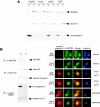
Localization of caspase 3 and AKAP95. (A) Localization of AKAP95 and procaspase 3 in normal cells. HepG2, HeLa, Jurkat, and 293T cells were harvested and fractionated into pellet (P) and supernatant (S) fractions after lysis with digitonin. Each fraction was subjected to SDS-PAGE and immunoblotted with anti-AKAP95 monoclonal antibody (upper panel), anti-lamin B1 polyclonal antibodies as a nuclear fraction marker (middle panel), or anti-caspase 3 monoclonal antibody (lower panel). (B) Colocalization of active caspase 3 and AKAP95 in apoptotic cells. HepG2 cells treated with an agonistic anti-Fas antibody in the presence of actinomycin D for 12 h were fractionated as described for panel A. Each fraction was subjected to SDS-PAGE and immunoblotted with anti-AKAP95 monoclonal antibody, anti-lamin B1 polyclonal antibodies, or anti-caspase 3 polyclonal antibodies (antibody 9662; Cell Signaling Technology) that detect both procaspase 3 and caspase 3-p17 as indicated (left panel). HepG2 cells were treated without or with an agonistic anti-Fas antibody in the presence of actinomycin D for 12 h or with etoposide for 40 h. After fixation and permeabilization, the cells were incubated with anti-active caspase 3 polyclonal (antibody 2622) or monoclonal (CS-1) antibodies, with anti-AKAP95 monoclonal or polyclonal antibodies, and with Texas Red (TXRD)- or fluorescein isothiocyanate-labeled secondary antibodies, followed by staining the nuclei with Hoechst 33342 (right panel).
FIG. 5.
FIG. 5.
Function of AKAP95 in nuclear morphological changes of apoptotic cells. (A) Diagram showing the NLS mutations of AKAP95. Mutated amino acid residues are underlined. NMTS, nuclear matrix targeting site; RII, type II PKA regulatory subunit. (B) Localization of GFP-AKAP95 fusion proteins with or without mutations. 293T cells were transiently transfected with GFP or GFP-AKAP95 expression plasmids as indicated. After 18 h, cells were observed by fluorescence microscopy. (C) Amino acid sequences of active caspase 3 binding region in AKAP95. Conserved EQT sequence between human and mouse AKAP95 is underlined. (D and E) Overexpression of AKAP95 with NLS mutations inhibits nuclear morphological changes. HepG2 cells were transfected with GFP-AKAP95 expression plasmids as indicated. After 24 h, cells were treated with an agonistic anti-Fas antibody in the presence of actinomycin D for 12 h (D) or 15 h (E) and collected and fixed with 3.7% formaldehyde for 10 min. After staining with Hoechst 33342, the percentage of the cells showing normal nuclei to the total transfected cells was measured. At least 100 GFP-positive cells were counted for each measurement in all experiments. The data (mean ± the standard deviation) were obtained from at least three independent experiments. Significant test results (P values) are shown. *, P < 0.05; **, P < 0.01; ***, P < 0.001.

Similar articles

Cited by

References

    1. Akileswaran, L., J. W. Taraska, J. A. Sayer, J. M. Gettemy, and V. M. Coghlan. 2001. A-kinase-anchoring protein AKAP95 is targeted to the nuclear matrix and associates with p68 RNA helicase. J. Biol. Chem. 276:17448-17454. - PubMed
    1. Alnemri, E. S., D. J. Livingston, D. W. Nicholson, G. Salvesen, N. A. Thornberry, W. W. Wong, and J. Yuan. 1996. Human ICE/CED-3 protease nomenclature. Cell 87:171. - PubMed
    1. Baird, G. S., D. A. Zacharias, and R. Y. Tsien. 2000. Biochemistry, mutagenesis, and oligomerization of DsRed, a red fluorescent protein from coral. Proc. Natl. Acad. Sci. USA 97:11984-11989. - PMC - PubMed
    1. Chai, J., E. Shiozaki, S. M. Srinivasula, Q. Wu, P. Dataa, E. S. Alnemri, and Y. Shi. 2001. Structural basis of caspase-7 inhibition by XIAP. Cell 104: 769-780. - PubMed
    1. Chai, J., Q. Wu, E. Shiozaki, S. M. Srinivasula, E. S. Alnemri, and Y. Shi. 2001. Crystal structure of a procaspase-7 zymogen: mechanisms of activation and substrate binding. Cell 107:399-407. - PubMed

Publication types

MeSH terms