. 2005 Nov;25(21):9608-20.
doi: 10.1128/MCB.25.21.9608-9620.2005.

DNA damage-induced phosphorylation of MdmX at serine 367 activates p53 by targeting MdmX for Mdm2-dependent degradation

Affiliations

DNA damage-induced phosphorylation of MdmX at serine 367 activates p53 by targeting MdmX for Mdm2-dependent degradation

Koji Okamoto et al. Mol Cell Biol. 2005 Nov.

Abstract

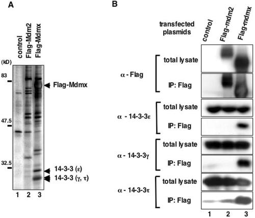
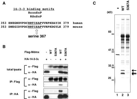
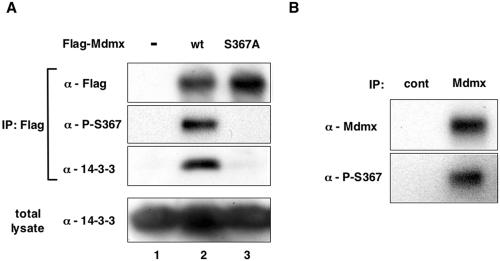
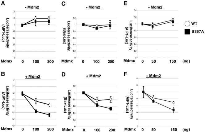
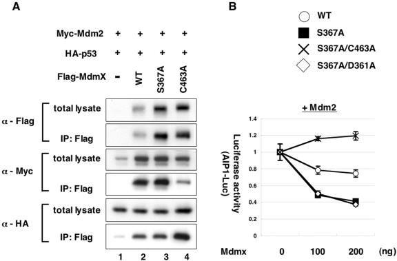
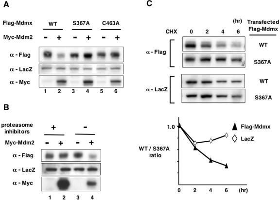
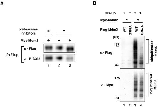
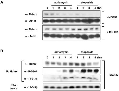
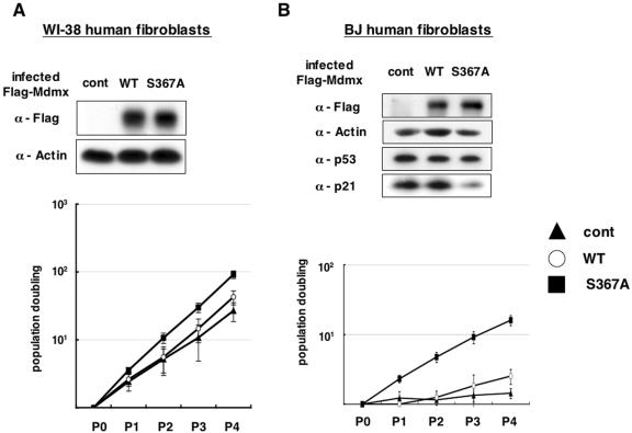
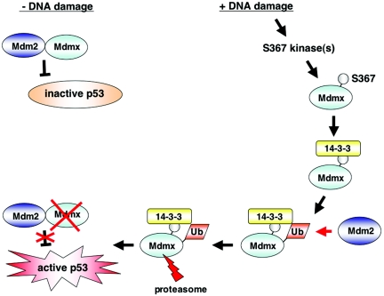
Understanding how p53 activity is regulated is crucial in elucidating mechanisms of cellular defense against cancer. Genetic data indicate that Mdmx as well as Mdm2 plays a major role in maintaining p53 activity at low levels in nonstressed cells. However, biochemical mechanisms of how Mdmx regulates p53 activity are not well understood. Through identification of Mdmx-binding proteins, we found that 14-3-3 proteins are associated with Mdmx. Mdmx harbors a consensus sequence for binding of 14-3-3. Serine 367 (S367) is located within the putative binding sequence for 14-3-3, and its substitution with alanine (S367A) abolishes binding of Mdmx to 14-3-3. Transfection assays indicated that the S367A mutation, in cooperation with Mdm2, enhances the ability of Mdmx to repress the transcriptional activity of p53. The S367A mutant is more resistant to Mdm2-dependent ubiquitination and degradation than wild-type Mdmx, and Mdmx phosphorylated at S367 is preferentially degraded by Mdm2. Several types of DNA damage markedly enhance S367 phosphorylation, coinciding with increased binding of Mdmx to 14-3-3 and accelerated Mdmx degradation. Furthermore, promotion of growth of normal human fibroblasts after introduction of Mdmx is enhanced by the S367 mutation. We propose that Mdmx phosphorylation at S367 plays an important role in p53 activation after DNA damage by triggering Mdm2-dependent degradation of Mdmx.


Figures

FIG. 1.
FIG. 1.
14-3-3 proteins bind to Mdmx. (A) Flag-Mdmx, Flag-Mdm2, or a control vector was transfected into COS-1 cells, and lysates prepared from transfected cells were used to purify Flag-tagged proteins on anti-Flag affinity columns as described in Materials and Methods. Polypeptides copurified with Flag-tagged proteins were separated on an SDS-PAGE gel and visualized by silver staining. (B) Polypeptides copurified with Flag-tagged proteins as described for panel A were separated on an SDS-PAGE gel, and Western blot analyses were performed with anti-Flag antibody (M2), or with anti-14-3-3 antibodies which detect the indicated isoforms.
FIG. 2.
FIG. 2.
Alanine substitution at S367 of Mdmx abolishes binding of 14-3-3 proteins to Mdmx. (A) Mdmx harbors an amino acid sequence that matches known 14-3-3 binding motifs. Previously reported consensus sequences for 14-3-3 binding sites are shown in the upper part of the figure (RXXXSXP and RSXSXP) (9), and aligned with amino acid sequences from human and mouse Mdmx. Amino acid sequences that match 14-3-3 binding motifs are underlined. The putative phosphorylation site (serine 367) is indicated by an arrow. (B) Wild-type Flag-Mdmx, the Flag-S367A mutant Mdmx, or the control vector was transfected into H1299 cells alone or together with HA-14-3-3ɛ. Lysates from transfected cells were used to immunoprecipitate Flag-Mdmx proteins with anti-Flag antibody or HA-14-3-3ɛ proteins with anti-HA antibody. The anti-Flag immunoprecipitates, the anti-HA immunoprecipitates, or total lysates were then analyzed by Western blot analyses with anti-Flag antibody or anti-HA antibody. (C) The control vector, wild-type Flag-Mdmx, or the S367A mutant was transfected into COS-1 cells, and lysates from transfected cells were used to purify Flag-Mdmx on agarose conjugated with anti-Flag antibody as described for Fig. 1A. Proteins copurified with Flag-Mdmx were visualized by silver staining. Polypeptides corresponding to the 14-3-3 proteins are shown by arrows.
FIG. 3.
FIG. 3.
S367 of Mdmx is phosphorylated in vivo. (A) WI-38 cells were infected with the control viruses or retroviruses that express wild-type Flag-Mdmx or the S367A mutant. After selection of infected cells by hygromycin resistance, lysates from infected cells were used for immunoprecipitation (IP) with anti-Flag antibody. Total lysates or theanti-Flag immunoprecipitates were then analyzed by Western blot analyses with anti-Flag antibody, anti-phospho-S367 antibody, or an anti-14-3-3 antibody that detects all isoforms (K-19). (B) Lysates from MCF-7 cells were immunoprecipitated with anti-Mdmx antibody (D-19; Santa-Cruz) and analyzed by Western blot analyses with a mixture of monoclonal anti-Mdmx antibodies (see Materials and Methods) or anti-phospho-S367 antibody.
FIG. 4.
FIG. 4.
Cooperative repression of p53 transcriptional activity by Mdmx and Mdm2 is augmented by the S367A mutation. H1299 cells (A, B, C, and D) or p53−/−/Mdmx−/− MEFs (E and F) were transfected with HA-p53, Renilla luciferase control vector, and firefly luciferase vector linked to the aip1 (A, B, E, and F) or the bax (C and D) promoter, together with (B, D, and F) or without (A, C, and E) Myc-Mdm2. Increasing amounts of wild-type Flag-Mdmx or the S367A mutant were cotransfected as indicated at the bottom of each graph. In experiments with Mdm2 cotransfection (B, D, and F), 100 ng (B and D) or 200 ng (F) of Myc-Mdm2 was introduced. In all experiments, 50 ng of HA-p53 was transfected and the total amount of DNA was adjusted to 2 μg with pBluescript plasmid (Stratagene); 24 h after transfection, cells were lysed and luciferase activity was measured using the dual-luciferase assay system (Promega). Mean values (±standard deviation) from three independent experiments were determined. Basal promoter activity expressed in the absence of HA-p53 was measured and subtracted in each experiment. Values presented were calculated as follows: value from cells transfected with indicated amount of Mdmx/value from cells transfected without Mdmx.
FIG. 5.
FIG. 5.
Characterization of the Mdmx mutants. (A) H1299 cells were transfected with 100 ng of Myc-Mdm2 and 50 ng of HA-p53 together with 200 ng of wild-type Flag-Mdmx, the indicated Flag-Mdmx mutant, or the control vector. Lysates from transfected cells were used for immunoprecipitation (IP) with anti-Flag antibody, and the anti-Flag immunoprecipitates or total lysates were analyzed by Western blot analyses with anti-Flag antibody, anti-Myc antibody, or anti-HA antibody. (B) H1299 cells were cotransfected as described for Fig. 4. Increasing amounts of wild-type Flag-Mdmx or indicated Mdmx mutant were transfected together with Myc-Mdm2, HA-p53, Renilla luciferase control vector, and firefly luciferase vector linked to the aip1 promoter. Normalized values are calculated from luciferase activity and presented as described for Fig. 4.
FIG. 6.
FIG. 6.
S367A is resistant to Mdm2-mediated degradation of Mdmx. (A) We transfected 200 ng of wild-type Flag-Mdmx or its mutants (S367A or C463A) into H1299 cells together with the LacZ expression vector (pCH110), with or without 100 ng of Myc-Mdm2; 24 h after transfection, lysates prepared from transfected cells were used for Western blot analyses with anti-Flag antibody, anti-LacZ antibody, or anti-Myc antibody. (B) Wild-type Flag-Mdmx was transfected into H1299 cells together with the LacZ expression vector, with or without Myc-Mdm2 as described for panel A; 24 h after transfection, cells were incubated with proteasome inhibitors (30 μM ALLN, 50 μM MG132, plus 50 μM LLnL) or dimethyl sulfoxide (DMSO) for an additional 5 h. Western blot analyses were performed as described for panel A. (C) S367A mutation stabilizes Mdmx in the presence of Mdm2. (Upper panel) Wild-type Flag-Mdmx or the S367A mutant was transfected into H1299 cells together with the LacZ expression vector and Myc-Mdm2 as described for Fig. 6A; 24 h after transfection, 50 μg/ml of cycloheximide (CHX) was added to the medium, and cells were harvested at the indicated times. Lysates prepared from cycloheximide-treated cells were used for Western blot analyses with anti-Flag antibody or anti-LacZ antibody. (Bottom panel) Levels of introduced LacZ and Mdmx shown in the upper panel were quantified, and the wild-type/S367A ratio (values from cells transfected with wild-type Mdmx/values from cells transfected with the S367A mutant) was calculated at each time point. Each calculated value was normalized such that the wild-type/S367A ratio at 0 h is presented as 1.0.
FIG. 7.
FIG. 7.
S367 phosphorylation facilitates Mdm2-dependent degradation of Mdmx, and the S367A mutant is resistant to Mdm2-mediated ubiquitination. (A) Mdmx phosphorylated at S367 is preferentially degraded by Mdmx. Wild-type Flag-Mdmx was transfected into H1299 cells alone or together with Myc-Mdm2 as described for Fig. 6B; 24 h after transfection, cells were incubated with proteasome inhibitors as described for Fig. 6 or dimethyl sulfoxide for an additional 5 h. Subsequently, lysates prepared from transfected cells were used to immunoprecipitate Flag-Mdmx with anti-Flag antibody. Approximately equal amounts of immunoprecipitated Flag-Mdmx were used for Western blot analyses with anti-Flag antibody or anti-phospho-S367 antibody. (B) Either wild-type Flag-Mdmx or the S367A mutant was transfected into H1299 cells together with (His)6-tagged ubiquitin (His-Ub), with or without Myc-Mdm2; 24 h after transfection, cells were incubated with a proteasome inhibitor (50 μM MG132) for an additional 3 h. Subsequently, cells were lysed with a buffer containing 8 M urea, and lysates were used to purify His-ubiquitin on Ni-NTA-agarose (QIAGEN). Flag-Mdmx or Myc-Mdm2 conjugated with His-ubiquitin was detected by Western blot analyses with anti-Flag antibody or anti-Myc antibody, respectively.
FIG. 8.
FIG. 8.
Induction of S367 phosphorylation after DNA damage is associated with increased binding of 14-3-3 to Mdmx and accelerated Mdmx degradation. (A) MCF-7 cells were preincubated with dimethyl sulfoxide (−MG132) or 20 μM MG132 (+MG132) for 1 hour and then incubated with 3 μM adriamycin or 20 μM etoposide for the indicated periods. Subsequently lysates prepared from the drug-treated cells were used for Western blot analyses with anti-Mdmx antibody (D-19; Santa-Cruz) or anti-actin antibody. (B) MCF cells were preincubated with MG132 and exposed to DNA-damaging compounds as described in A. Cell lysates prepared from the drug-treated cells were used for immunoprecipitation with anti-Mdmx antibody (D-19). The anti-Mdmx immunoprecipitates or total lysates were used for Western blot analyses with the anti-Mdmx antibody (D-19), the anti-P-S367 antibody, or anti-14-3-3β antibody (Santa Cruz).
FIG. 9.
FIG. 9.
Introduction of the S367A mutant into normal human fibroblasts causes accelerated cell growth. Young WI-38 cells (A) and presenescent BJ cells (B) were infected with retroviruses that express wild-type Flag-Mdmx (circles), the S367A mutant (squares), or the control retroviruses (triangles). After selection of infected cells by hygromycin resistance, cells were grown under the 3T3 protocol. Cells were split every 3 days, and cell numbers were calculated and plotted on a logarithmic scale. Cell counting started at the end of drug selection. Mean values (±standard deviation) from three independent experiments are presented to evaluate growth of the infected cells. The accumulated number of population doublings is shown on a log scale. Lysates were prepared from infected cells after drug selection, and Western blot analyses were performed with anti-Flag antibody, antiactin antibody, anti-p53 antibody, and anti-p21 antibody.
FIG. 10.
FIG. 10.
Model of the molecular mechanism of p53 activation by S367 phosphorylation. In cells with low levels of activity of the S367 kinase(s), Mdm2 and Mdmx cooperatively repress transcriptional activity of p53. After DNA damage induces activation of the S367 kinase(s) and S367 phosphorylation, 14-3-3 binds to Mdmx. Binding of 14-3-3 to Mdmx induces ubiquitination of Mdmx by Mdm2. Ubiquitination of Mdmx causes its proteasomal degradation and leads to p53 activation.

Similar articles

Cited by

References

    1. Appella, E., and C. W. Anderson. 2001. Post-translational modifications and activation of p53 by genotoxic stresses. Eur. J. Biochem. 268:2764-2772. - PubMed
    1. Atadja, P., H. Wong, I. Garkavtsev, C. Veillette, and K. Riabowol. 1995. Increased activity of p53 in senescing fibroblasts. Proc. Natl. Acad. Sci. USA 92:8348-8352. - PMC - PubMed
    1. Badciong, J. C., and A. L. Haas. 2002. MdmX is a RING finger ubiquitin ligase capable of synergistically enhancing Mdm2 ubiquitination. J. Biol. Chem. 277:49668-49675. - PubMed
    1. Banin, S., L. Moyal, S. Shieh, Y. Taya, C. W. Anderson, L. Chessa, N. I. Smorodinsky, C. Prives, Y. Reiss, Y. Shiloh, and Y. Ziv. 1998. Enhanced phosphorylation of p53 by ATM in response to DNA damage. Science 281:1674-1677. - PubMed
    1. Barak, Y., T. Juven, R. Haffner, and M. Oren. 1993. mdm2 expression is induced by wild type p53 activity. EMBO J. 12:461-468. - PMC - PubMed

Publication types

MeSH terms