. 2005 Nov;25(21):9632-45.
doi: 10.1128/MCB.25.21.9632-9645.2005.

Met/Hepatocyte growth factor receptor ubiquitination suppresses transformation and is required for Hrs phosphorylation

Affiliations

Met/Hepatocyte growth factor receptor ubiquitination suppresses transformation and is required for Hrs phosphorylation

Jasmine V Abella et al. Mol Cell Biol. 2005 Nov.

Abstract

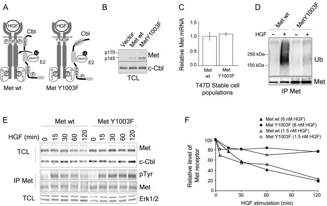
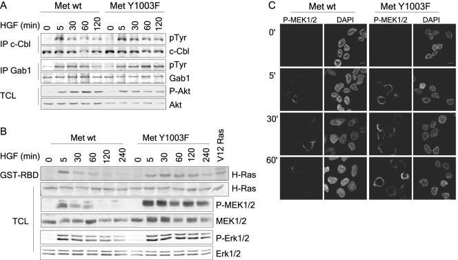
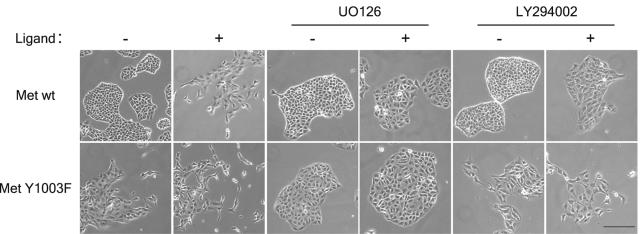
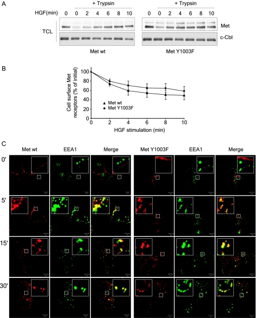
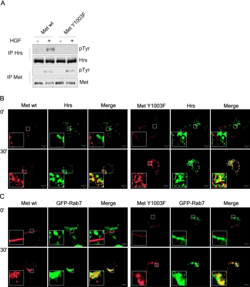
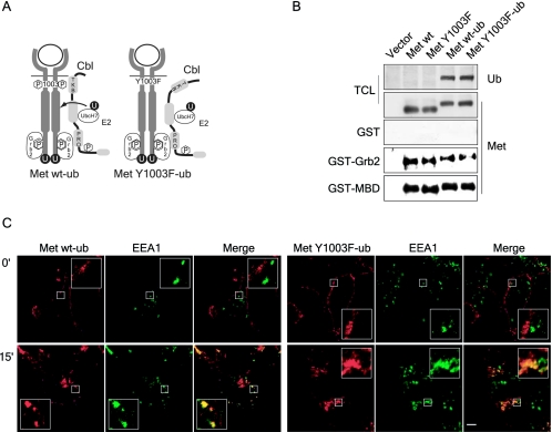
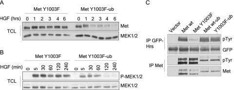
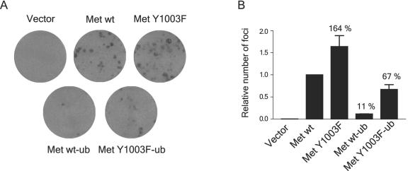
The Met receptor tyrosine kinase (RTK) regulates epithelial remodeling, dispersal, and invasion and is deregulated in many human cancers. It is now accepted that impaired down-regulation, as well as sustained activation, of RTKs could contribute to their deregulation. Down-regulation of the Met receptor involves ligand-induced internalization, ubiquitination by Cbl ubiquitin ligases, and lysosomal degradation. Here we report that a ubiquitination-deficient Met receptor mutant (Y1003F) is tumorigenic in vivo. The Met Y1003F mutant is internalized, and undergoes endosomal trafficking with kinetics similar to the wild-type Met receptor, yet is inefficiently targeted for degradation. This results in sustained activation of Met Y1003F and downstream signals involving the Ras-mitogen-activated protein kinase pathway, cell transformation, and tumorigenesis. Although Met Y1003F undergoes endosomal trafficking and localizes with the cargo-sorting protein Hrs, it is unable to induce phosphorylation of Hrs. Fusion of monoubiquitin to Met Y1003F is sufficient to decrease Met receptor stability and prevent sustained MEK1/2 activation. In addition, this rescues Hrs tyrosine phosphorylation and decreases transformation in a focus-forming assay. These results demonstrate that Cbl-dependent ubiquitination is dispensable for Met internalization but is critical to target the Met receptor to components of the lysosomal sorting machinery and to suppress its inherent transforming activity.


Figures

FIG. 1.
FIG. 1.
The Met Y1003F receptor mutant is poorly ubiquitinated and its degradation is delayed upon HGF stimulation. (A) Schematic representation of Cbl recruitment to the Met receptor. The Y1003F substitution abrogates binding of the Cbl TKB domain to the Met receptor. The Met Y1003F receptor mutant still recruits and tyrosine phosphorylates Cbl via the Grb2 adaptor. (B) Expression levels of the Met wild-type and Y1003F receptors in retrovirally infected stable T47D cell populations. After selection with neomycin for 2 weeks, cells expressing Met at their cell surface were sorted using FACS. Total cell lysates (TCL) were blotted with Met (antibody 144) and c-Cbl antibodies. The p170 band represents the uncleaved Met precursor and the p145 band represents the processed α chain of the receptor. (C) Met RNA expression levels from T47D cell populations were determined using quantitative real-time PCR. The graph represents the mean ± standard deviation of triplicate samples. (D) T47D cells were stimulated with 3 nM HGF for 2 min and lysed immediately in boiling 2% SDS. Lysates were boiled for 10 min, diluted to 0.4% SDS, 2% Triton and then Met receptor proteins were immunoprecipitated with antibody 144 and blotted with ubiquitin antibodies, stripped and reblotted with Met antibodies. (E) T47D cells were stimulated with either 6 nM HGF (top two panels) or 1.5 nM HGF (bottom three panels) for the indicated amount of time. Lysates were treated as indicated. (F) Met protein levels were quantified using the ImageJ 1.63 software and were corrected using Cbl (6 nM HGF) and Erk2 (1.5 nM HGF) protein levels.
FIG. 2.
FIG. 2.
NIH 3T3 fibroblast cells expressing the ubiquitination-deficient Met Y1003F receptor are tumorigenic. (A) Met Y1003F-expressing cells have an altered morphology. Phase contrast pictures of T47D cells unstimulated or stimulated for 24 h with 1.5 nM HGF. (B) NIH 3T3 cell populations expressing either wild-type Met or Met Y1003F were injected subcutaneously into nude mice. The results represent the mean tumor volume obtained from eight measurements and are representative of two independent experiments. (C) Pictures of representative tumors as well as Met protein expression in the NIH 3T3 cell populations injected and in excised tumors. The tumors were lysed in TGH buffer and total cell lysates (TCL) were blotted with Met (DL-21) antibodies. Then, the membrane was stained with Coomassie brilliant blue.
FIG. 3.
FIG. 3.
Activation of the Ras-MAPK but not the PI3K pathway is sustained downstream from Met Y1003F. (A) T47D cells were stimulated with 1.5 nM HGF for the indicated times and lysed in TGH buffer. In the upper two panels, c-Cbl proteins were immunoprecipitated and immunoblotted with phosphotyrosine antibodies. Membranes were stripped and blotted with Cbl antibodies. In the middle two panels, Gab1 proteins were immunoprecipitated and blotted with both antiphosphotyrosine, 4G10, and Gab1 antibodies using the Odyssey Infrared Imaging System (LI-COR). In the bottom two panels, lysates were blotted with either phospho-Ser473 or total Akt antibodies as indicated using the Odyssey system. (B) T47D cells were stimulated with 1.5 nM HGF and lysed as above. Upper two panels: Ras activation was determined using an in vitro binding assay with the GST-RBD fusion protein, with HEK293T cells expressing V12 H-Ras as a positive control. Panels were blotted with an H-Ras antibody using the Odyssey System. In the bottom four panels, cell lysates were blotted with phospho-Ser217/221 MEK1/2, total MEK1/2, phospho-Thr202/Tyr204 Erk1/2, and total Erk1/2 antibodies as indicated using the Odyssey system. (C) T47D cells were plated on coverslips, serum starved for 16 h, and stimulated with 1.5 nM HGF at 37°C for the indicated time points. Coverslips were fixed in 3% paraformaldehyde (PFA), and stained with phospho-Ser217/221 MEK1/2 (first panel). Cell nuclei were visualized using 4′,6′-diamidino-2-phenylindole (DAPI) (second panel). Confocal images were taken with a 100× objective and 2× zoom. The bar represents 5 μm.
FIG. 4.
FIG. 4.
Ras-MAPK is required for Met Y1003F increased biological activity. MDCK cells stably expressing either wild-type CSF-Met or CSF-Met Y1003F, where indicated, were treated with CSF (2.7 nM), UO126 (20 μM) and LY294002 (25 μM) for 24 h. After 24 h, phase-contrast pictures of live cells were taken with a Zeiss Axiovision 135 microscope under a 10× objective. The bar represents 100 μm.
FIG. 5.
FIG. 5.
Ubiquitination-deficient Met receptor mutant is internalized. (A) T47D cells were stimulated with 3 nM HGF at 37°C and then transferred on ice. Cells were acidified for 10 min and then treated with trypsin for 30 min. Trypsin was inhibited before cells were lysed. Lysates were blotted with Met (DL-21) and c-Cbl antibodies. (B) T47D cells were stimulated with 3 nM HGF at 37°C and then transferred on ice. Cells were acidified for 10 min and then labeled with Met AF276 antibody as described under Materials and Methods. The mean fluorescence intensity per cell was measured by flow cytometry and the percentage of Met receptors remaining at the cell surface over time is plotted. The graph represents the mean ± standard deviation of three independent experiments. (C) Both wild-type Met and Met Y1003F internalize and traffic to early endosomes. T47D cells were plated on coverslips, and 16 h later stimulated with 1.5 nM HGF at 37°C for the indicated times. Coverslips were fixed in 3% PFA and stained with Met AF276 (red) and EEA1 (green) antibodies. Confocal images were taken with a 100× objective and 2× zoom. Yellow staining represents colocalization between Met and EEA1. The bar represents 5 μm.
FIG. 6.
FIG. 6.
Ubiquitination-deficient Met receptor mutant is unable to induce Hrs tyrosine phosphorylation. (A) T47D cells were serum starved for 16 h and stimulated where indicated with 1.5 nM HGF for 8 min. Cells were then lysed in TGH buffer and Hrs and Met proteins were immunoprecipitated and first blotted with phosphotyrosine antibodies, stripped, and reblotted with Hrs and Met antibodies as indicated. (B and C) Wild-type Met and Met Y1003F both colocalize with endogenous Hrs and transfected GFP-Rab7. T47D cells, nontransfected (B) and transfected (C), were plated on coverslips and 16 h later stimulated with 1.5 nM HGF at 37°C for the indicated times. Coverslips were fixed with 3% PFA and stained with (B and C) Met AF276 (red) and (B) Hrs (green) antibodies. Yellow staining represents colocalization between Met and Hrs or Met and GFP-Rab7. Confocal images were taken with a 100× objective and 2× zoom. The bar represents 5 μm.
FIG. 7.
FIG. 7.
Fusion of monoubiquitin to the carboxy terminus of the Met receptor does not alter its maturation to the cell surface, recruitment of signaling proteins and its HGF-induced internalization. (A) Schematic representation of ubiquitinated wild-type and Y1003 Met chimeric receptors. Met receptor chimeras were generated with a single ubiquitin moiety fused to the carboxy-terminal end of the full-length wild-type and Y1003F Met receptors. The seven lysine residues within the ubiquitin moiety were replaced with arginine residues to prevent polyubiquitination and the carboxy-terminal glycine residue was replaced with a valine residue to prevent conjugation to free amino groups. (B) The chimeric receptors are properly expressed and tyrosine phosphorylated. HEK 293 cells were transiently transfected with the indicated CSF-Met constructs. In the top two panels, lysates were blotted with Met (antibody 144) and ubiquitin antibodies. In the bottom three panels, lysates were incubated with GST alone, GST-Grb2, or GST-MBD (Met binding domain of Gab1). In vitro binding assays and total cell lysates (TCL) were immunoblotted for Met. (C) T47D cells were plated on coverslips and 16 h later stimulated with 1.5 nM HGF at 37°C for 15 min. Coverslips were fixed in 3% PFA and stained with Met AF276 (red) and EEA1 (green) antibodies. Confocal images were taken with a 100× objective and 2× zoom. Yellow staining represents colocalization between Met and EEA1. The bar represents 5 μm.
FIG. 8.
FIG. 8.
Monoubiquitination of the Met Y1003F receptor leads to its degradation and restores HGF-induced Hrs tyrosine phosphorylation. (A) T47D cells treated with 100 μg/ml cycloheximide were stimulated with 3 nM HGF for the indicated amount of time. Total cell lysates (TCL) (30 μg) were immunoblotted for Met (DL-21) and Erk. (B) T47D cells were stimulated with 1.5 nM HGF for the indicated times and lysed in TGH buffer. Cell lysates were blotted with phospho-Ser217/221 MEK1/2 and total MEK1/2. (C) HEK293 cells were cotransfected with GFP-Hrs and vector or the indicated CSF-Met constructs. GFP-Hrs and Met proteins were immunoprecipitated and blotted with phosphotyrosine antibodies, then stripped and reblotted for GFP and Met, respectively.
FIG. 9.
FIG. 9.
Monoubiquitination of the Met Y1003F receptor mutant is sufficient to reverse cell transformation. Rat-1 fibroblast cells were infected with retroviruses encoding the various CSF-Met constructs and grown for 3 weeks in the presence of 0.3 nM of colony-stimulating factor 1. (A) Representative pictures of cell monolayers. (B) The graph represents the relative number of foci per dish corrected for the PFU of each retrovirus, from two independent experiments.

References

    1. Bache, K. G., C. Raiborg, A. Mehlum, and H. Stenmark. 2003. STAM and Hrs are subunits of a multivalent ubiquitin-binding complex on early endosomes. J. Biol. Chem. 278:12513-12521. - PubMed
    1. Bache, K. G., T. Slagsvold, and H. Stenmark. 2004. Defective downregulation of receptor tyrosine kinases in cancer. EMBO J. 23:2707-2712. - PMC - PubMed
    1. Bao, J., G. Gur, and Y. Yarden. 2003. Src promotes destruction of c-Cbl: implications for oncogenic synergy between Src and growth factor receptors. Proc. Natl. Acad. Sci. USA 100:2438-2443. - PMC - PubMed
    1. Bilodeau, P. S., J. L. Urbanowski, S. C. Winistorfer, and R. C. Piper. 2002. The Vps27p Hse1p complex binds ubiquitin and mediates endosomal protein sorting. Nat. Cell Biol. 4:534-539. - PubMed
    1. Bilodeau, P. S., S. C. Winistorfer, W. R. Kearney, A. D. Robertson, and R. C. Piper. 2003. Vps27-Hse1 and ESCRT-I complexes cooperate to increase efficiency of sorting ubiquitinated proteins at the endosome. J. Cell Biol. 163:237-243. - PMC - PubMed

Publication types

MeSH terms

Substances

LinkOut - more resources