. 2005 Nov;25(21):9646-60.
doi: 10.1128/MCB.25.21.9646-9660.2005.

Impaired immune responses and prolonged allograft survival in Sly1 mutant mice

Affiliations

Impaired immune responses and prolonged allograft survival in Sly1 mutant mice

Sandra Beer et al. Mol Cell Biol. 2005 Nov.

Abstract

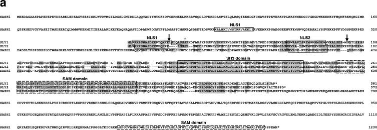
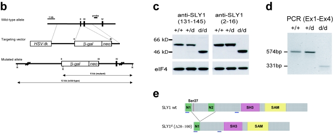
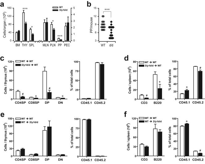
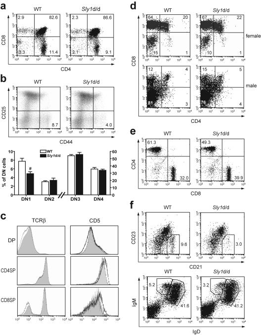
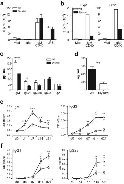
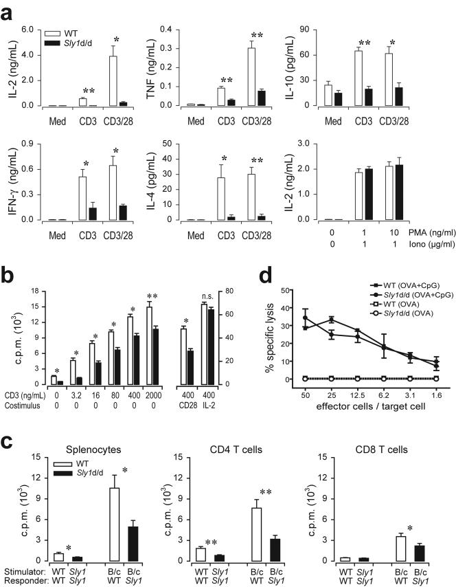
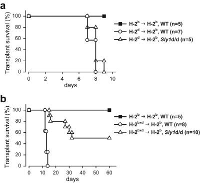
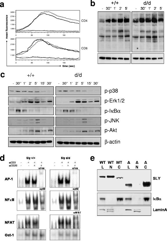
Adaptive immunity is crucial for protective host defense and the development of immunological disorders. SLY1 was recently identified as an X-chromosomal SH3 protein that is serine phosphorylated (Ser27) upon B-and T-cell receptor engagement. Here, we demonstrate that SLY1 is localized in the cytoplasm and the nucleus of immunocytes. We generated mice expressing a mutant version of SLY1 lacking Ser27 and a functional nuclear localization signal. The defective SLY1 (SLY1(d)) protein is localized exclusively in the cytoplasm. B- and T-cell proliferation is attenuated and T-cell cytokine production is severely reduced. Sly1(d/d) mice exhibit reduced lymphoid organ sizes, diminished marginal zone B-cell numbers, and severely impaired antibody responses against T-dependent and -independent antigens. Importantly, survival of semi-identical cardiac allografts was substantially prolonged in Sly1(d/d) mice. These results define SLY1 as an essential molecular component for the full activation of adaptive immunity.


Figures

FIG. 1.
FIG. 1.
Generation of Sly1 mutant mice. (a) Amino acid sequence alignment of murine SLY1, SLY2, and SASH1 proteins is shown. Identical residues are shaded and protein domains are indicated by boxes. The arrows assign the deletion in SLY1d caused by skipping of exons 2 and 3. The asterisk marks phosphorylated Ser27. (b) The organization of part of the murine Sly1 genomic locus (top), the linearized targeting vector (middle), and the targeted Sly1 allele (bottom) are shown. The β-galactosidase cDNA and the neomycin resistance cassette were inserted between codon 26 (exon 2) and codon 72 (exon 3) of the Sly1 gene. The β-galactosidase cDNA contains a polyadenylation signal. Exon sequences are represented by solid rectangles, intron sequences by a black line (R, EcoRI). (c) Lysates of splenocytes from wild-type (+/+), heterozygous (+/d), and homozygous mutant (d/d) mice were analyzed by Western blot with two independent rabbit polyclonal antisera directed against amino acids 2 to 16 or 131 to 145 of the wild-type SLY1 protein. Antisera specific for eIF4 served as loading control. (d) The skipping of exons 2 and 3 was revealed by reverse transcription-PCR with exon 1- and exon 4-specific primers in splenic mRNA from wild-type (+/+) and heterozygous (+/d) as well as homozygous (d/d) mutant mice. (e) Schematic representation of the mutant SLY1d protein. N, nuclear localization signal; blue lines, immunogenic peptides for generation of SLY1-specific antisera.
FIG. 1.
FIG. 1.
Generation of Sly1 mutant mice. (a) Amino acid sequence alignment of murine SLY1, SLY2, and SASH1 proteins is shown. Identical residues are shaded and protein domains are indicated by boxes. The arrows assign the deletion in SLY1d caused by skipping of exons 2 and 3. The asterisk marks phosphorylated Ser27. (b) The organization of part of the murine Sly1 genomic locus (top), the linearized targeting vector (middle), and the targeted Sly1 allele (bottom) are shown. The β-galactosidase cDNA and the neomycin resistance cassette were inserted between codon 26 (exon 2) and codon 72 (exon 3) of the Sly1 gene. The β-galactosidase cDNA contains a polyadenylation signal. Exon sequences are represented by solid rectangles, intron sequences by a black line (R, EcoRI). (c) Lysates of splenocytes from wild-type (+/+), heterozygous (+/d), and homozygous mutant (d/d) mice were analyzed by Western blot with two independent rabbit polyclonal antisera directed against amino acids 2 to 16 or 131 to 145 of the wild-type SLY1 protein. Antisera specific for eIF4 served as loading control. (d) The skipping of exons 2 and 3 was revealed by reverse transcription-PCR with exon 1- and exon 4-specific primers in splenic mRNA from wild-type (+/+) and heterozygous (+/d) as well as homozygous (d/d) mutant mice. (e) Schematic representation of the mutant SLY1d protein. N, nuclear localization signal; blue lines, immunogenic peptides for generation of SLY1-specific antisera.
FIG. 2.
FIG. 2.
Influence of the Sly1d/d mutation on lymphoid organogenesis. (a) Total cell numbers were determined from bone marrow (BM), thymus (THY), spleen (SPL), mesenteric (MLN) and peripheral (PLN) lymph nodes, Peyer's patches (PP), and in peritoneal lavage (PEC) of wild-type littermates (white bars) and Sly1 mutant mice (solid bars) with 18 mice analyzed in each group. (b) Numbers of macroscopically visible Peyer's patches were counted. Each symbol represents an individual mouse (WT, wild-type littermate controls; d/d, Sly1 mutant mice). (c and e) Chimeric mice were generated by adoptive transfer of bone marrow cells from wild-type or Sly1d/d mice (both CD45.2+) into lethally irradiated CD45.1+ wild-type recipients or (d and f) by injection of CD45.1+ wild-type bone marrow cells into CD45.2+ wild-type and Sly1d/d recipients. Recipient mice were analyzed 4 weeks after adoptive transfer for CD4/CD8-defined thymic subsets (c and e) and the presence of CD3+ and B220+ cells in spleen (d and f). Results of adoptive transfer experiments are derived from 4 or 5 mice per group. *, P < 0.05; #, P < 0.01; ***, P < 0.001 (Student's t test). DP, double-positive; DN, double-negative.
FIG. 3.
FIG. 3.
Development of T and B cells in Sly1d/d mice. (a) Thymocyte subsets were analyzed by flow cytometry using antibodies against CD4 and CD8. (b) Subsets of double-negative thymocytes were further differentiated by their CD44 and CD25 expression profile (upper panels). In the lower panel, results from eight to nine independent mice per group are summarized. (c) Histograms depict surface levels of T-cell receptor β(TCRβ) and CD5 on double-positive (DP) and single-positive (SP) thymocytes (wild type [WT], shaded graphs; Sly1d/d mice, black lines). (d) Sly1d/d mice were mated with mice expressing a transgenic T-cell receptor specific for the HY antigen in the context with H2-Db. Thymocytes were gated for HY-TCRhigh cells and the proportions of CD4- and CD8-defined subsets present in female and male mice are shown. (e) The proportions of CD4 and CD8 T cells (gated on CD3+ cells) and (f) CD21hi CD23lo (gated on IgM+ cells) or IgMhi IgDlo marginal zone B cells in spleens of wild-type and Sly1d/d mice were determined. All plots are representative of at least 6 independent mice per group. #, P < 0.01 (Student's t test).
FIG.4.
FIG.4.
B-cell activation and humoral immune responses in Sly1d/d mice. B220+ CD21int CD23hi follicular B cells (a) and B220+ CD21hi CD23lo marginal zone B cells (b) were purified from spleens of wild-type (white bars) and Sly1 mutant (solid bars) mice and were stimulated with anti-IgM and anti-CD40 or 40 ng/ml lipopolysaccharide (LPS). [3H]thymidine incorporation was measured after 3 days (n = 4 experiments). (c) Basal levels of immunoglobulin isotypes were determined in serum of untreated mice (n = 17 to 19 mice per group). (d) Levels of IgM in the peritoneal lavage (n = 8 to 9 mice per group). (e) For analysis of T-independent antibody responses, the time course of TNP-specific IgM and IgG3 antibody levels was determined in serum of wild-type (triangles) and Sly1 mutant (circles) mice immunized with TNP-Ficoll (n = 7 to 8 mice per group). (f) T-dependent production of TNP-specific antibody isotypes was measured after immunization of mice with TNP-CGG adsorbed to alum (n = 4 mice per group for day 4 and day 7; n = 8 mice per group for day 0, day 14, and day 21). *, P < 0.05; **, P < 0.01; ***, P < 0.001 (Student's t test).
FIG. 5.
FIG. 5.
Impaired antigen receptor-mediated activation of Sly1d/d T cells. (a) Splenocytes from wild-type (white bars) or Sly1d/d mice (solid bars) were stimulated with CD3 (2 μg/ml) alone or a combination of CD3 (0.4 μg/ml) and CD28 (2.5 μg/ml) antibodies and cytokine production was measured after 18 h. Additionally, IL-2 release was determined upon stimulation with the indicated concentrations of phorbol myristate acetate and ionomycin (n = 4 to 6 mice per group). (b) Splenocytes from wild-type (white bars) or Sly1d/d mice (solid bars) were stimulated by the addition of antibodies against CD3. CD28 antibodies (2.5 μg/ml) or IL-2 (10 ng/ml) were added as indicated. After 66 h [3H]thymidine was added and incorporation was measured after additional 16 h (n = 12 mice per group). (c) Total splenocytes or purified splenic CD4 and CD8 T cells were prepared from Sly1d/d or wild-type (WT) mice and stimulated with allogeneic BALB/c splenocytes (B/c) or autologous cells (n = 5 mice per group). (d) Mice were immunized with ovalbumin in the presence or absence of CpG DNA (CpG) as an adjuvant. Draining lymph node cells were harvested 4 days later and cultured in vitro with recombinant IL-2 for an additional 4 days. Cytotoxic T-cell activity was assayed using EL4 target cells pulsed with the SIINFEKL peptide (n = 4 independent experiments). *, P < 0.05, and **, P < 0.01 (Student's t test), for all experiments.
FIG. 6.
FIG. 6.
Improved cardiac allograft acceptance in Sly1d/d mice. Fully allogeneic H-2d (a) or semi-identical H-2bxd (b) cardiac allografts were transplanted into wild-type or Sly1 mutant mice. For control, wild-type mice received syngeneic H-2b grafts. Allograft survival was monitored for the indicated time periods.
FIG. 7.
FIG. 7.
Influence of the Sly1d/d mutation on T-cell receptor signaling and subcellular localization of SLY1 protein. (a) Splenocytes were loaded with Fluo-4 and stained for CD4 and CD8 on ice. After warming, baseline fluorescence was determined and T-cell receptor signaling was induced by CD3 cross-linking. (b) T cells were stimulated with anti-CD3 for the indicated periods. Cell lysates were analyzed by Western blotting with antibodies against phosphotyrosine, as well as (c) phospho-p38, phospho-Erk1/2, phospho-JNK, phospho-Akt, and phospho-IκBα as indicated. Blots were stripped and reprobed with anti-β-actin as a loading control. (d) T cells were stimulated for 16 h as indicated and mobility shift assays with specific oligonucleotides were performed. Antibodies used for supershifts are indicated. (e) For subcellular localization of SLY1 protein, whole-cell lysates (L) as well as cytoplasmic (C) and nuclear (N) fractions from lymphocytes were prepared, followed by Western blot analysis with anti-SLY1 (2-16) antibody. The blots were stripped and reprobed with anti-IκBα and anti-lamin A/C antibodies to control for the purity of the preparations.

References

    1. Alimzhanov, M. B., D. V. Kuprash, M. H. Kosco-Vilbois, A. Luz, R. L. Turetskaya, A. Tarakhovsky, K. Rajewsky, S. A. Nedospasov, and K. Pfeffer. 1997. Abnormal development of secondary lymphoid tissues in lymphotoxin β-deficient mice. Proc. Natl. Acad. Sci. USA 94:9302-9307. - PMC - PubMed
    1. Astoul, E., A. D. Laurence, N. Totty, S. Beer, D. R. Alexander, and D. A. Cantrell. 2003. Approaches to define antigen receptor induced serine kinase signal transduction pathways. J. Biol. Chem. 278:9267-9275. - PubMed
    1. Azzam, H. S., A. Grinberg, K. Lui, H. Shen, E. W. Shores, and P. E. Love. 1998. CD5 expression is developmentally regulated by T-cell receptor (TCR) signals and TCR avidity. J. Exp. Med. 188:2301-2311. - PMC - PubMed
    1. Beer, S., A. B. Simins, A. Schuster, and B. Holzmann. 2001. Molecular cloning and characterization of a novel SH3 protein (SLY) preferentially expressed in lymphoid cells. Biochim. Biophys. Acta 1520:89-93. - PubMed
    1. Cho, H. J., K. Takabayashi, P. M. Cheng, M. D. Nguyen, M. Corr, S. Tuck, and E. Raz. 2000. Immunostimulatory DNA-based vaccines induce cytotoxic lymphocyte activity by a T-helper cell-independent mechanism. Nat. Biotechnol. 18:509-514. - PubMed

Publication types

MeSH terms