Comparative Study
. 2005 Nov;11(11):1188-96.
doi: 10.1038/nm1307. Epub 2005 Oct 16.

Akt1 regulates pathological angiogenesis, vascular maturation and permeability in vivo

Affiliations
Comparative Study

Akt1 regulates pathological angiogenesis, vascular maturation and permeability in vivo

Juhua Chen et al. Nat Med. 2005 Nov.

Abstract

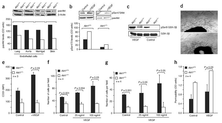
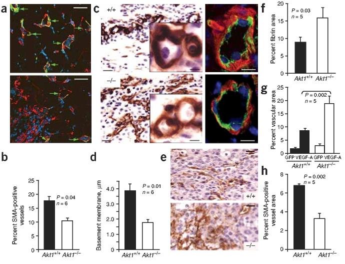
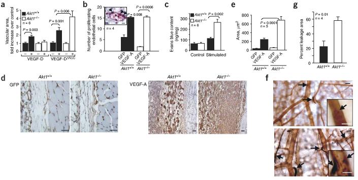
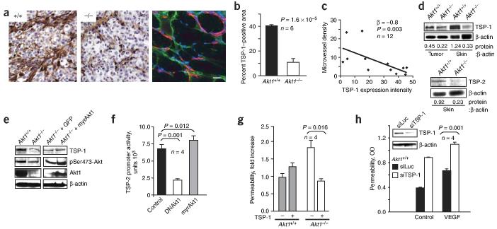
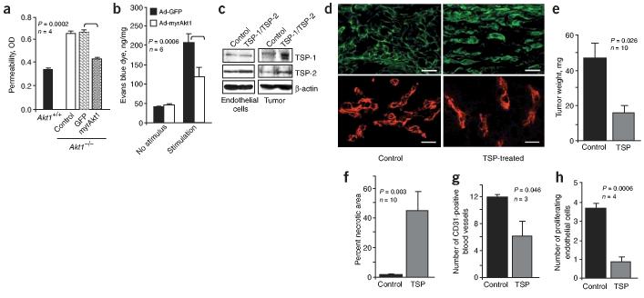
Akt kinases control essential cellular functions, including proliferation, apoptosis, metabolism and transcription, and have been proposed as promising targets for treatment of angiogenesis-dependent pathologies, such as cancer and ischemic injury. But their precise roles in neovascularization remain elusive. Here we show that Akt1 is the predominant isoform in vascular cells and describe the unexpected consequences of Akt1 knockout on vascular integrity and pathological angiogenesis. Angiogenic responses in three distinct in vivo models were enhanced in Akt1(-/-) mice; these enhanced responses were associated with impairment of blood vessel maturation and increased vascular permeability. Although impaired vascular maturation in Akt1(-/-) mice may be attributed to reduced activation of endothelial nitric oxide synthase (eNOS), the major phenotypic changes in vascular permeability and angiogenesis were linked to reduced expression of two endogenous vascular regulators, thrombospondins 1 (TSP-1) and 2 (TSP-2). Re-expression of TSP-1 and TSP-2 in mice transplanted with wild-type bone marrow corrected the angiogenic abnormalities in Akt1(-/-) mice. These findings establish a crucial role of an Akt-thrombospondin axis in angiogenesis.


Figures

Figure 1
Figure 1
Deficiency of Akt1, the predominant Akt isoform in endothelial cells, impairs their function ex vivo. (a) Western blot analyses and band densitometry of total Akt expression (using panAkt antibodies) in endothelial cells isolated from lungs, aorta and Matrigel plugs and in skin of wild-type (filled bars) and Akt1/ (open bars) mice. (b) Western blot analyses of total phosphorylated Akt in lung endothelial cells from wild-type and Akt1/ mice in response to 20 ng/ml VEGF. (c) Western blot analyses of phosphorylated GSK-3β in lung endothelial cells from wild-type and Akt1/ mice in the presence and absence of VEGF (20 ng/ml). (d) Micrographs of representative aortic ring microvessels from wild-type (top) and Akt1/ (bottom) animals grown in the presence of VEGF. Scale bars, 20 μm. (e) Quantification of vascular outgrowth in the presence or absence of VEGF (30 ng/ml) was performed by FACS analysis using CD31-specific antibodies. MFI, mean of fluorescence intensity. (f,g) Endothelial cell migration toward vitronectin (f) and fibronectin (g) was stimulated by 25 and 100 ng/ml of VEGF as indicated. (h) Permeability of endothelial monolayers in vitro. Wild-type and Akt1/ endothelial cell monolayers were grown in transwells for at least 1 week to reach confluency and then stimulated with VEGF (30 ng/ml) or were unstimulated. n refers to number of times each experiment was repeated for each condition.
Figure 2
Figure 2
Enhanced in vivo angiogenesis in Akt1/ mice. (a,b) Angiogenesis in Matrigel implants stimulated with VEGF. (a) Hemoglobin content in Matrigel implants from Akt1/ mice was higher compared to that from wild-type mice. Inset shows the microscopic appearance of 5-d-old Matrigel implants with 60 ng/ml VEGF from wild-type (top) and Akt1/ (bottom) mice (original magnification, ×2). (b) Increased endothelial cell infiltration into Matrigel in Akt1/ versus wild-type mice. (ce) Angiogenesis induced by implanted tumors. (c) Comparison of CD31 staining of tumors grown in wild-type (left) and Akt1/ mice (right). Scale bars, 20 μm. (d) Image analysis shows the increased density of blood vessels (the number of vessels per tissue area) in tumors grown in Akt1/ versus wild-type mice. (e) Comparison of vascular area in tumors implanted in wild-type and Akt1/ mice. (f) A representative photograph of the necrotic area in the center of tumor grown in wild-type mice (left). No necrosis was observed in tumors from Akt1/ mice (right). Sections were stained with hematoxylin and eosin. Scale bars, 50 μm. (g) Increased number of CD105-positive blood vessels (green fluorescence) in frozen sections of tumors grown in Akt1/ mice (middle) compared to wild-type mice (left). Right panel shows costaining for CD105 (green) and laminin (red) in the tumor blood vessels in wild-type mice. Nuclei are stained with DAPI. Scale bars, 10 μm. (h) Quantitative analysis of CD105-positive vessels in tumors from Akt1/ mice compared to wild-type mice. n refers to the number of mice in each group.
Figure 3
Figure 3
Neovasculature in Akt1−/− mice is immature and leaky. Immunohistochemistry and image analysis of tumors implanted into wild-type and Akt1−/− mice. (a) Costaining for SMA (green) and CD31 (red) in tumors from wild-type and Akt1−/− mice. Nuclei are stained with DAPI. Arrowheads indicate SMA-positive vascular structures. Scale bars, 50 μm. (b) Quantification of the data presented in a. (c) Laminin-stained blood vessels in tumors grown in wild-type (top) and Akt1−/− mice (bottom). Insets show higher-magnification images. Scale bars, 20 μm. Inset scale bar, 5 μm. Immunohistochemistry (right panels) shows laminin (red), CD105 (green) and nuclei (blue). Scale bars, 10 μm. (d) Thickness of laminin-positive basement membrane in microvessels formed in wild-type and in Akt1−/− mice. (e) Fibrin deposition (brown) in tumors grown in wild-type (top) and Akt1−/− (bottom) mice. Scale bars, 20 μm. (f) Quantification of fibrin-positive area shown in e. (g,h) Angiogenesis induced by VEGF-A165 or GFP (control) adenoviruses. (g) Comparison of vascular area in vWF-stained sections of wild-type and Akt1−/− skin upon stimulation with VEGF-A165. Representative images are in Supplementary Figure 2 online. (h) Comparison of SMA-positive vascular area in wild-type and Akt1−/− skin 7 d after injection of Ad-VEGF-A. Representative images are in Supplementary Fig. 2 online.
Figure 4
Figure 4
Characterization of vascular responses in Akt1−/− mice. (a) Comparison of angiogenesis induced by full-length VEGF-D and VEGF-DΔNΔC in wild-type and Akt1−/− mice. (b) Comparison of endothelial cell proliferation in Ad-GFP– or Ad-VEGF-A–treated skin of wild-type and Akt1−/− mice based on Ki-67 staining. Inset shows a representative image of proliferating endothelial cells (arrowheads) in a skin blood vessel. Scale bar, 10 μm. (c) Plasma leakage in ear skin assessed by Evans blue extravasation after treatment with mustard oil, with no difference in control (unstimulated) ears. (d) Vascular permeability in wild-type and Akt1−/− mice in response to Ad-VEGF-A treatment. Fibrin staining shows increased leakage of fibrin in Akt1−/− compared to wild-type mice. Scale bars, 50 μm. (e) Area of fibrin deposition in skins from wild-type and Akt1−/− mice shown in d. (f) Comparison of ear microvessel morphology and leakage sites in ricin-stained whole mounts of ear skin in wild-type and Akt1−/− mice upon stimulation with mustard oil. Dark patches (shown by arrows) indicate sites of exposed basement membrane. There was an increased number of dark patches in blood vessels in Akt1−/− (lower panel, inset) compared to wild-type mice (upper panel, inset). Scale bars, 50 μm. Inset scale bar, 20 μm. (g) Quantitative analysis of the leakage area of wild-type and Akt1−/− blood vessels shown in f. n refers to the number of mice in each group.
Figure 5
Figure 5
Akt1 deficiency results in reduction of TSP levels. (a) TSP-1 staining in tumors from wild-type (left) and Akt1−/− mice (middle). Right panel shows costaining for TSP-1 (green) and CD31 (red) in tumors from Akt1−/− mice. Scale bar, 20 μm. (b) Comparison of TSP-1 staining in tumors from wild-type and Akt1−/− mice. (c) A reciprocal correlation existed between vascularity and TSP-1 expression in tumors. (d) Western blot and subsequent densitometry shows reduced levels of TSP-1 and TSP-2 in tissues from Akt1−/−compared to wild-type mice. (e) Levels of TSP-1, phosphorylated Akt, Akt1 and β-actin in wild-type and Akt1−/− endothelial cells upon treatment with Ad-myrAkt1 or Ad-GFP determined by western blot. There were increased levels of TSP-1 upon re-expression of Akt1. (f) Luciferase activity measured in HUVECs cotransfected with a Tsp 2 promoter/luciferase construct and retroviral constructs encoding DNAKt1, myrAkt1 or control. (g) Permeability of endothelial cell monolayers from wild-type and Akt1−/− mice in the presence or absence of TSP-1. Exogenous TSP-1 (1 μg/ml) decreased the permeability of Akt1−/− endothelial cell monolayers, whereas it slightly increased the permeability of wild-type endothelial cell monolayers. (h) The consequences of downregulation of TSP-1 by siRNA on the permeability of endothelial cell monolayers. Endothelial cells were treated with TSP-1 siRNA lentivirus (siTSP-1) or luciferase siRNA (siLuc) as a control. Inset shows TSP-1 levels after siRNA treatment. The permeability of endothelial cells was stimulated by 30 ng/ml VEGF-A165 as indicated. n refers to number of times each experiment was repeated for each condition.
Figure 6
Figure 6
An Akt1-TSP axis regulates vascular permeability and angiogenesis. (a) Comparison of the endothelial cell permeability of wild-type and Akt1−/− endothelial cells after treatment with Ad-myrAkt1 or Ad-GFP. Restoration of Akt resulted in the decrease of endothelial cell permeability to the level observed in wild-type endothelial cells. (b) Effect of prolonged Ad-myrAkt1 treatment on vascular permeability in Akt1−/− mice. Vascular leakage of Evans blue was stimulated by mustard oil (n = 6 mice per group). (c–h) TSP re-expression corrects the angiogenic phenotype of Akt1−/− mice. Primary endothelial cells and fibroblasts transfected with TSP-1 and TSP-2 were subcutaneously injected into Akt1−/− chimeric mice reconstituted with wild-type bone marrow followed by implantation of a mixture of tumor cells and TSP-overexpressing cells (Supplementary Methods online). (c) Western blot analysis shows increased expression of TSP-1 and TSP-2 in vitro in primary endothelial cells and fibroblasts cotransfected with plasmids carrying genes encoding TSP-1 and TSP-2. Increased TSP-1 and TSP-2 expression was also seen in the tumors grown in vivo. (d) Increased levels of TSP-1 (green staining, upper panels) and a decreased number of CD31-stained blood vessels (red, lower panels) in tumors upon re-expression of TSP-1 and TSP-2. Scale bar, 20 μm. (e) Effect of TSP-1 and TSP-2 on tumor weight. (f) Area of necrosis in tumors upon re-expression of TSP-1 and TSP-2. (g) Vascular density upon expression of TSP-1 and TSP-2 in tumors shown in d. (h) Expression of TSP-1 and TSP-2 resulted in decreased endothelial cell proliferation in vivo based on Ki67 staining (Fig. 4).

Similar articles

Cited by

References

    1. Risau W. Mechanisms of angiogenesis. Nature. 1997;386:671–674. - PubMed
    1. Folkman J. Angiogenesis in cancer, vascular, rheumatoid and other disease. Nat. Med. 1995;1:27–31. - PubMed
    1. Carmeliet P. Angiogenesis in health and disease. Nat. Med. 2003;9:653–660. - PubMed
    1. Jiang BH, Zheng JZ, Aoki M, Vogt PK. Phosphatidylinositol 3-kinase signaling mediates angiogenesis and expression of vascular endothelial growth factor in endothelial cells. Proc. Natl. Acad. Sci. USA. 2000;97:1749–1753. - PMC - PubMed
    1. Kandel ES, Hay N. The regulation and activities of the multifunctional serine/threonine kinase Akt/PKB. Exp. Cell Res. 1999;253:210–229. - PubMed

Publication types

MeSH terms